


Virtual Microscopy
|
|
||||
|
Virtual Microscopy |
||||
Case 1:
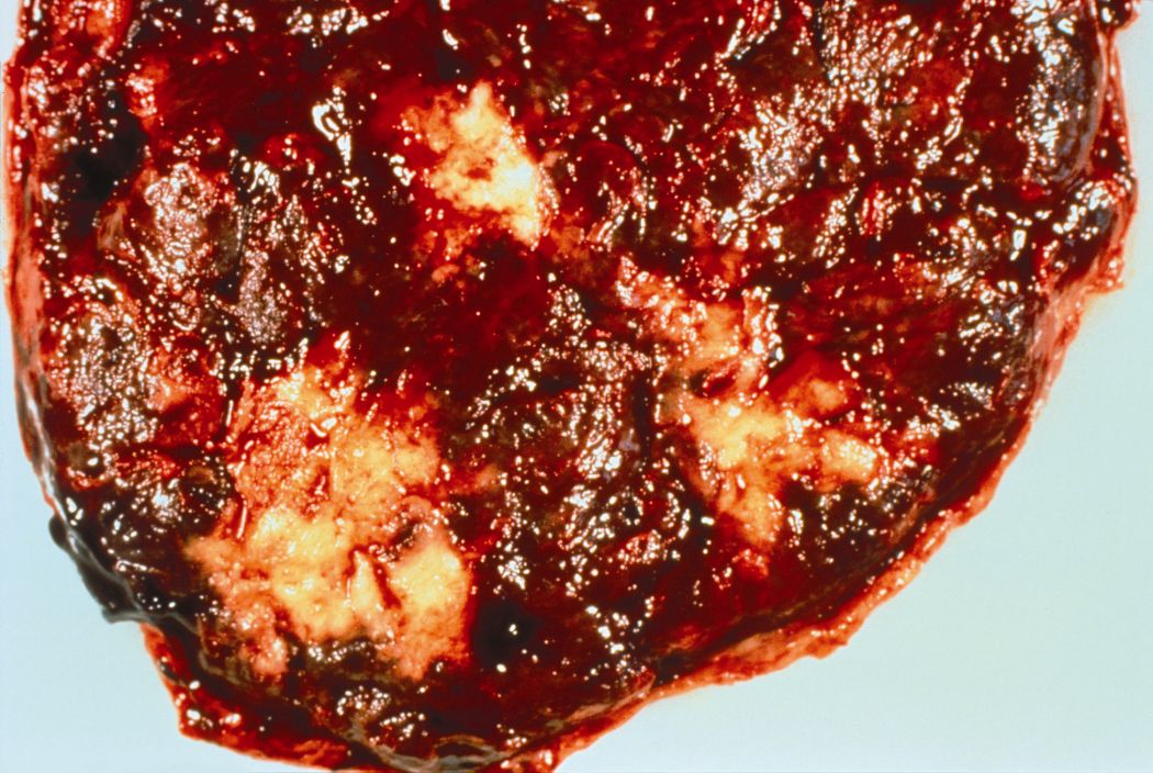
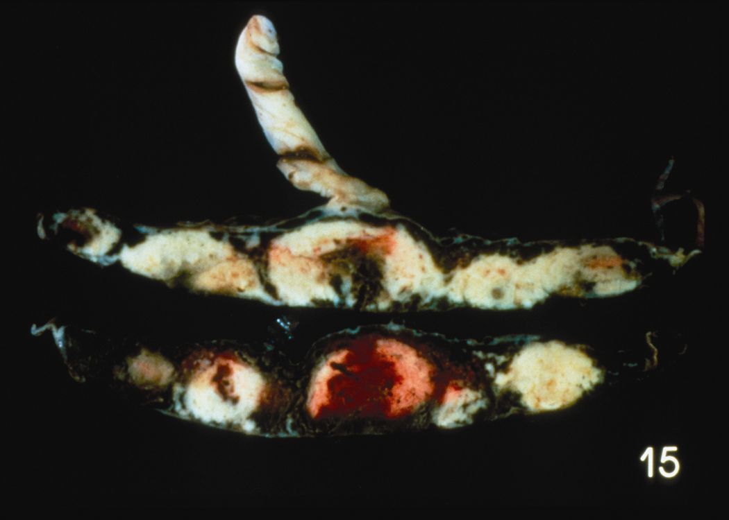
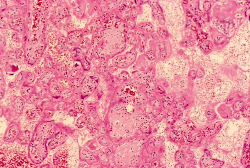
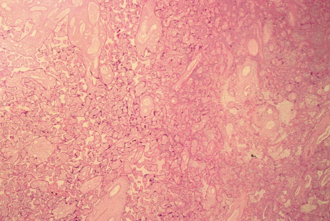
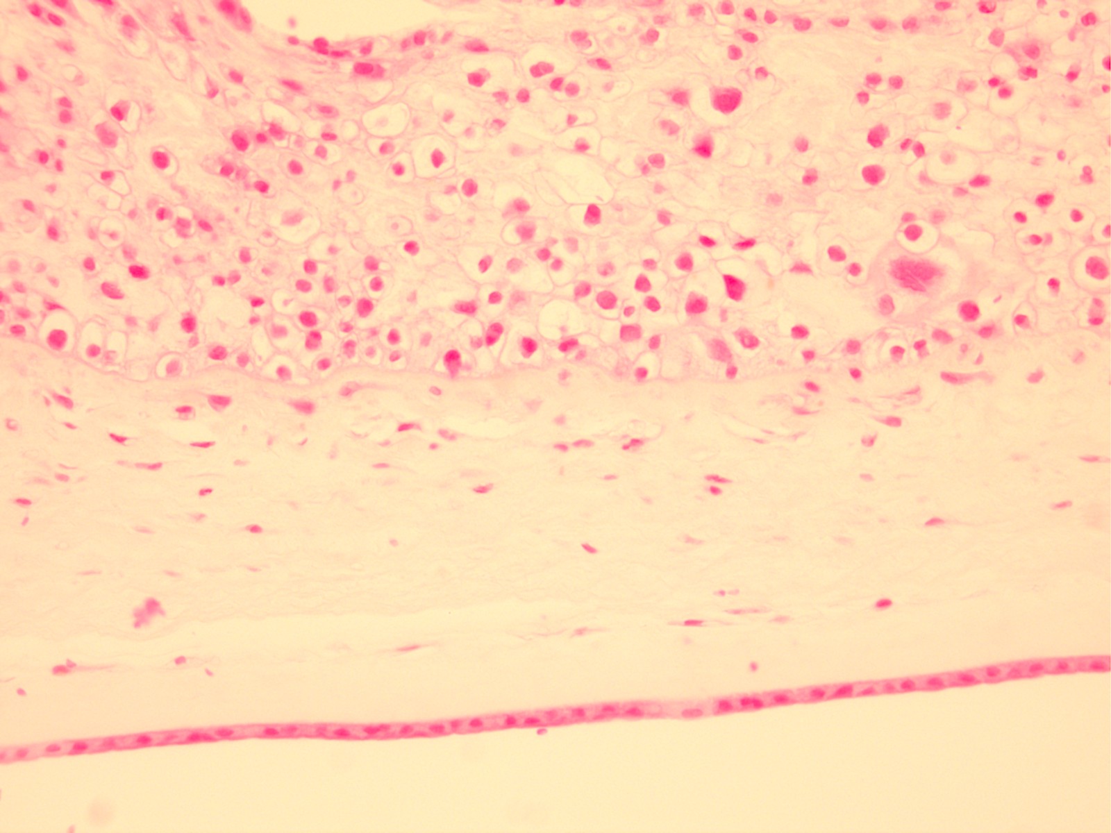
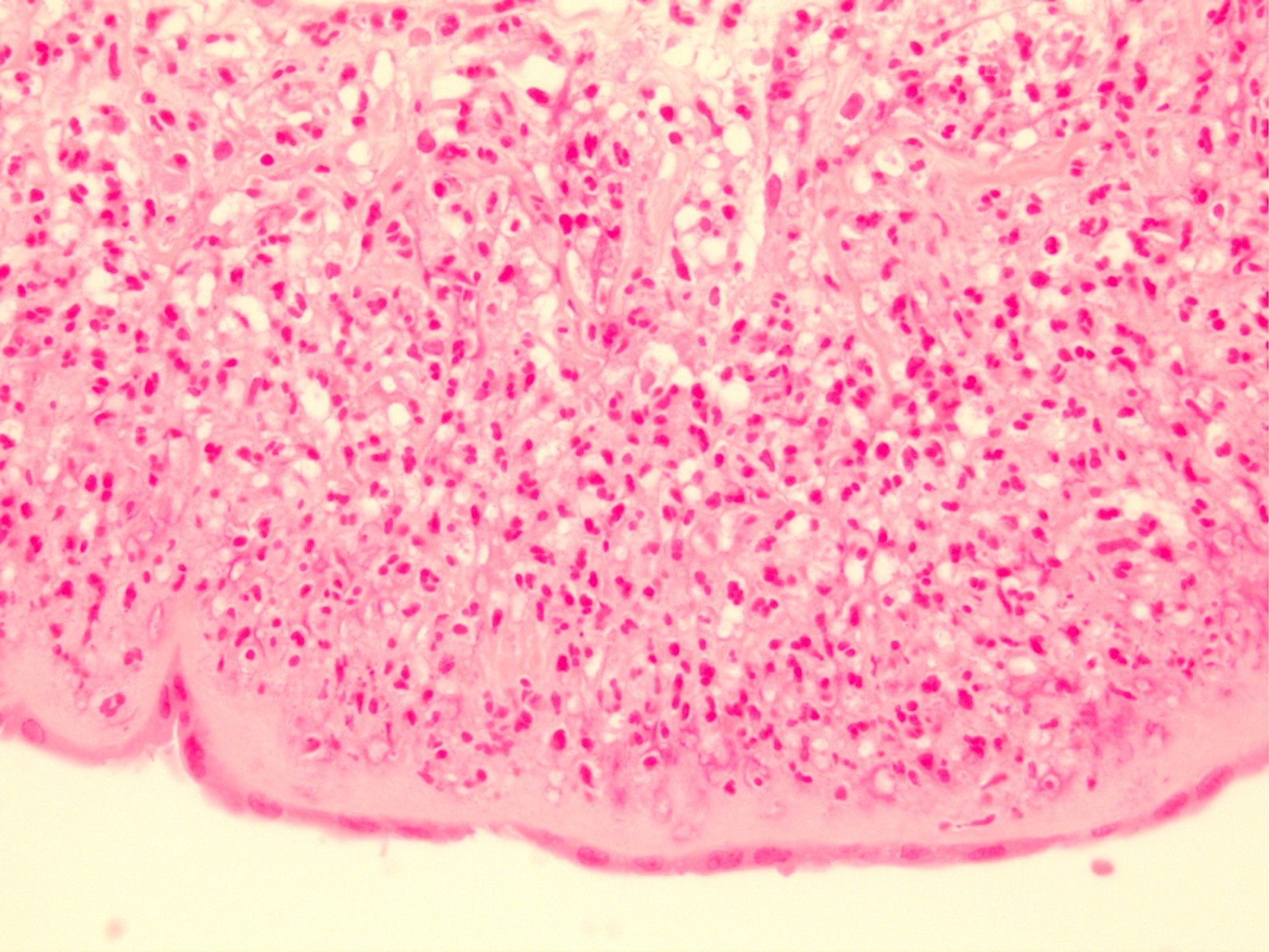
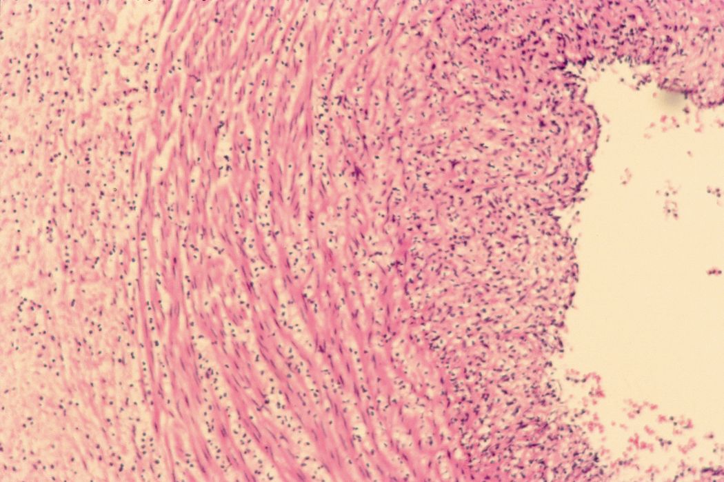
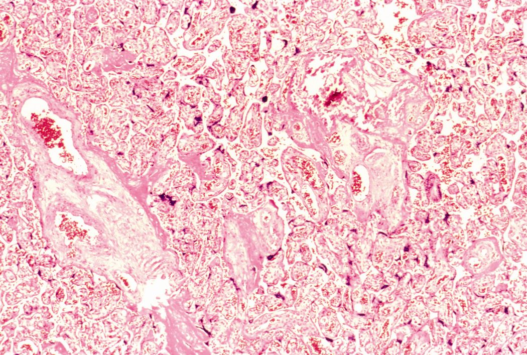
A thirty-year-old woman presents at 34 weeks gestation for a routine OB appointment. On examination she has an elevated blood pressure and had protein detected in her urine. Following labor induction at 36 weeks she delivered a healthy 7 pound 3-ounce female infant. Examination of the placenta revealed the following: [infarct – gross] [infarct – gross] [infarct – microscopic] [infarct – microscopic]
Case 2:
A 26 year old woman delivers a 6 pound 8 ounce male infant at 34 weeks gestation following premature rupture of membranes. The infant spikes a tempretature within several hours following delivery. [Normal placental membranes] [This baby’s placental membrane] [This baby’s umbilical cord]
Case 3:
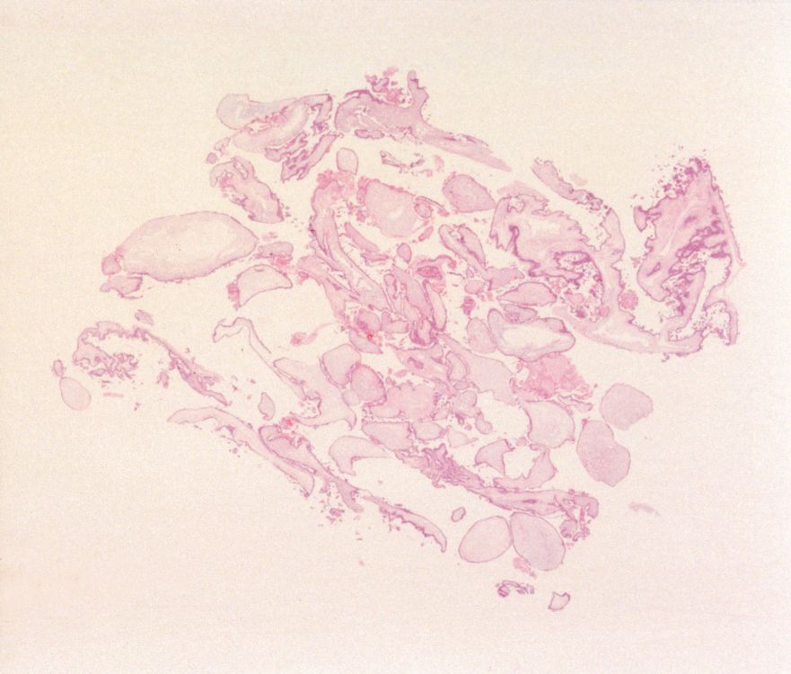
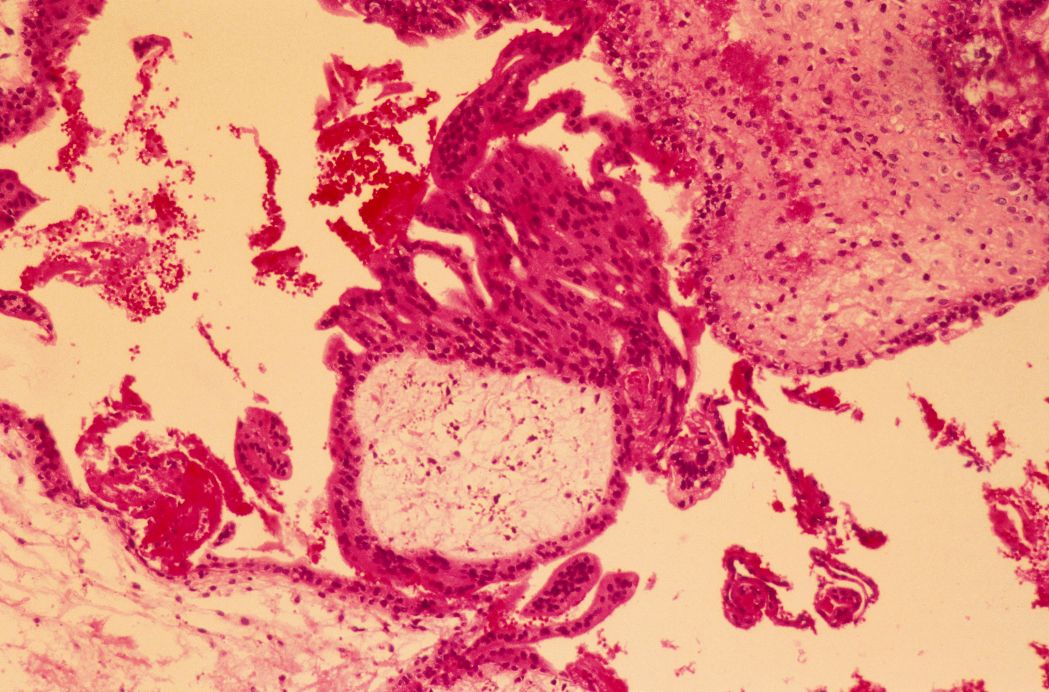
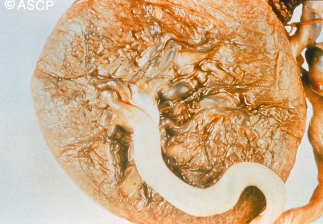
A 42 year old pregnant woman presents with vaginal bleeding at 9 weeks. On physical examination, the uterus was enlarged and fetal activity was not detected on ultrasound. [complete mole] [complete mole – gross] [complete mole]
Show and Tell: