


Virtual Microscopy
|
|
||||
|
Virtual Microscopy |
||||
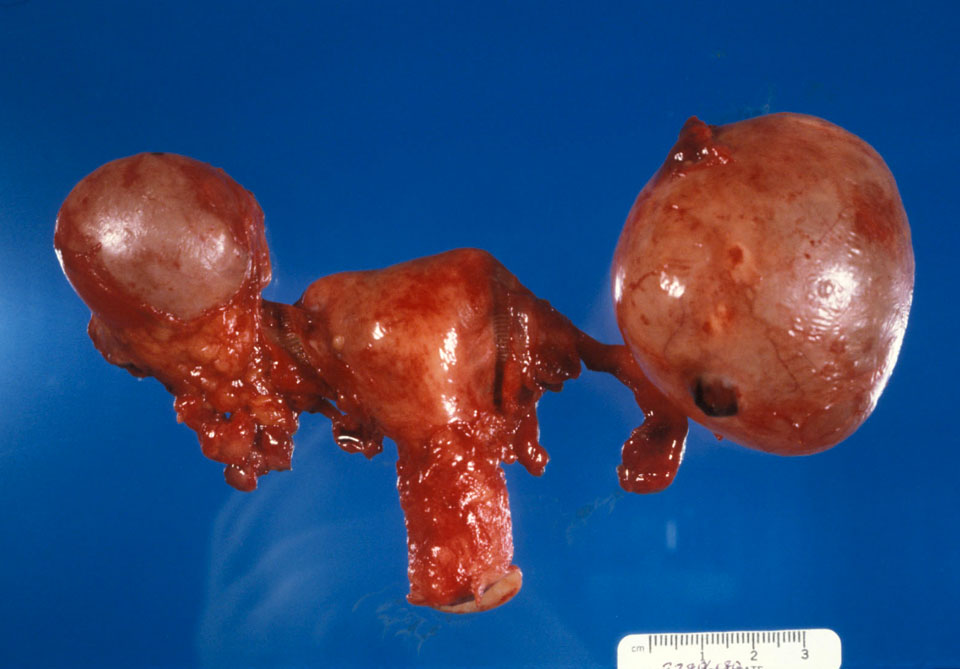
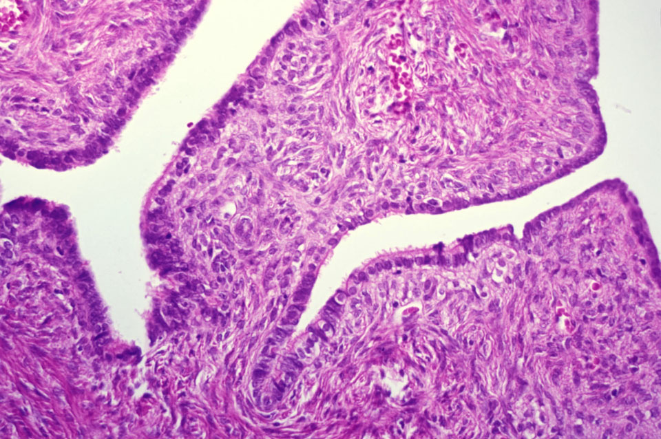
Case 1:
A 43 year old female was found to have a mass involving the left adnexa on a routine gynecologic exam. Transvaginal ultrasound showed a simple cystic mass. At surgery, a mass measuring 8 cm in diameter was found on the surface of the left ovary. [gross-opened benign cyst, hysterectomy/BSO, serous cystadenoma, gross of cystic tumor, mucinous cystadenoma]
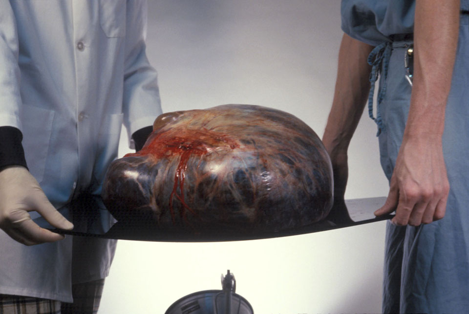
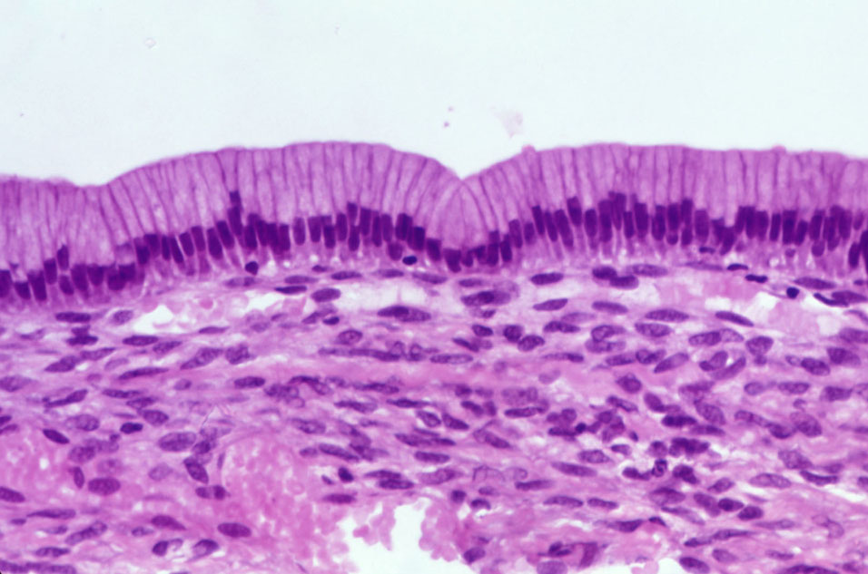
Case 2:
A 59 year old female presented with vague complaints of abdominal pain and increasing girth. An ultrasound showed bilateral complex solid/cystic adnexal masses, and she was immediately referred for surgical intervention. [serous cystadenocarcinoma]
Case 3:
A 29 year female was found to have a cystic mass on her right ovary during a C-section. A frozen section was requested. The pathologist called in to the operating room and said that the opened ovary contained a mass of hair and was partially ossified. A representative slide was reported as benign. [teratoma]