


Virtual Microscopy
|
|
||||
|
Virtual Microscopy |
||||
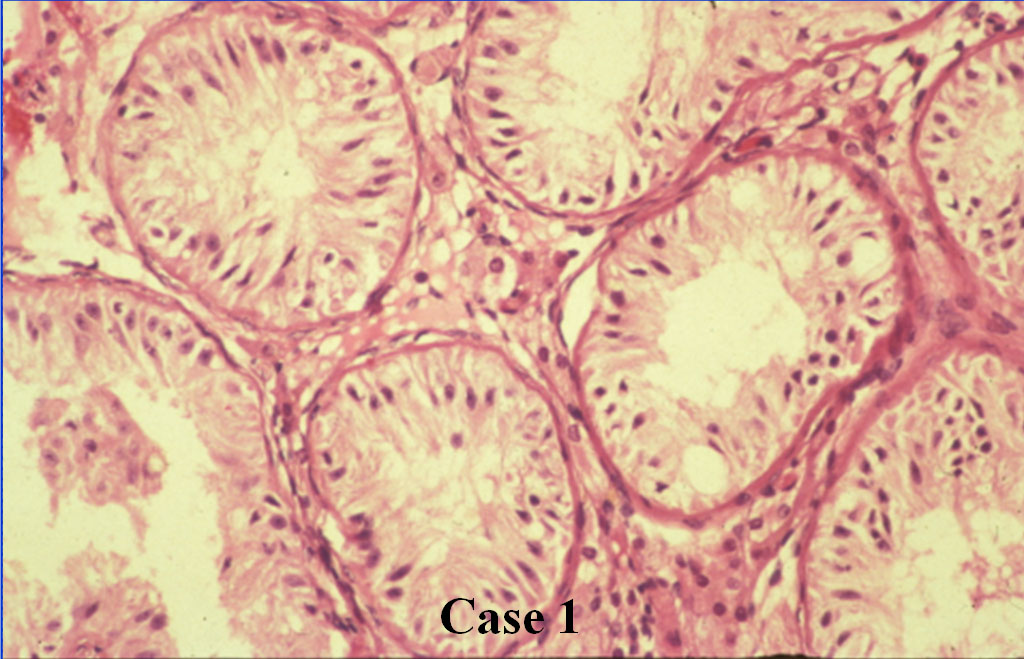
Case 1: Germinal Cell Aplasia
A 30-year-old male and his wife had a history of infertility. The examination of the wife was unremarkable. The patient had small testes. A testis biopsy was performed.
Photomicrograph: [biopsy]
Case 2: Benign Prostatic Hypertrophy
A 61 year old man developed increased frequency of urination, nocturia, urgency, and hesitancy.
Photomicrograph: [prostate 1] [prostate 2]
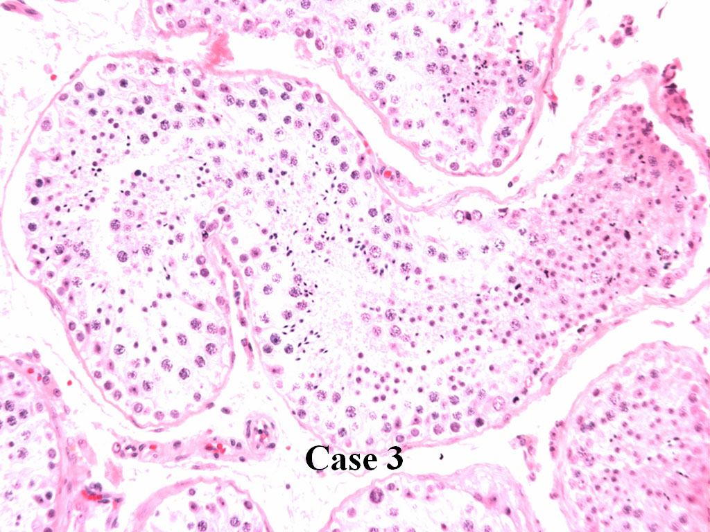
Case 3
A 35-year-old male and his wife had a two-year history of infertility. The examination of the wife yielded no functional or anatomic abnormalities. The patient had a semen analysis that contained no spermatozoa. A testis biopsy was performed.
Photomicrograph: [biopsy] [testis]