Virtual Microscopy > Reproductive > Pathology of Common Breast Lesions
Case 1:
A 63 year old female with a negative previous mammogram 5 years ago presents for routine screening. Suspicious radiodensities are noted which were not present on the previous films.
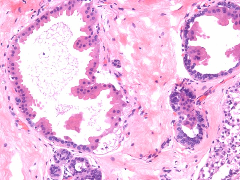
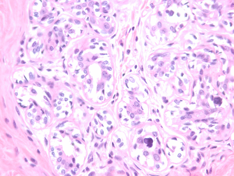
- What are the histologic findings of non-proliferative vs. proliferative fibrocystic changes? [apocrine metaplasia, ductal epithelial hyperplasia, microcalcifications, breast parenchyma]
- What is the clinical significance of these two different classes of fibrocystic changes?
Case 2:
A 35 year old female with a strong positive family history of breast cancer present with the request for BRCA testing to determine her risk for this disease.
- What is the significance of finding this mutation?
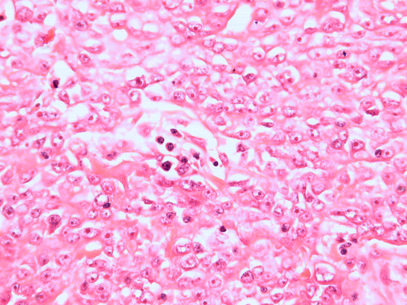
- What histology type of tumor corresponds to the BRCA1 gene type? [basal phenotype breast carcinoma #1, basal phenotype breast carcinoma #2]
- What histology type of tumor corresponds to the BRCA2 gene type? [ductal carcinoma of breast]
Case 3:
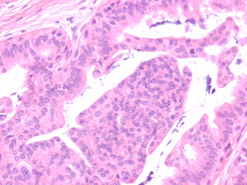
A 57 year old female with abnormal microcalcifications seen on routine mammogram was found to have DCIS (ductal carcinoma in situ) on core needle biopsy. [DCIS]. She was referred to a surgeon for needle localization biopsy which showed a 5 mm infiltrating ductal carcinoma in association with the DCIS. [ductal carcinoma of breast]
- What histologic changes distinguish DCIS from infiltrating ductal carcinoma? [DCIS and infiltrating carcinoma]
- What is the difference between DCIS and LCIS (lobular carcinoma in situ)? [DCIS 10X, DCIS 40X, DCIS and normal 10X, DCIS 40X, LCIS 40X]
- What is the clinical significance of infiltrating ductal vs. lobular carcinoma? [LCIS & inv. lob.]
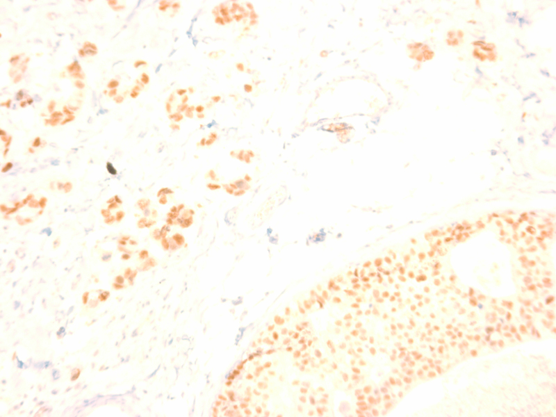
- Why are estrogen and progesterone receptor analyses performed routinely on tumor tissue? [er. pos. DCIS]
Show and Tell:
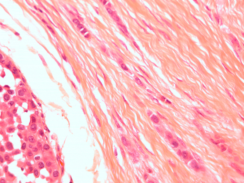
[Fibroadenoma #1]
[Fibroadenoma #2]
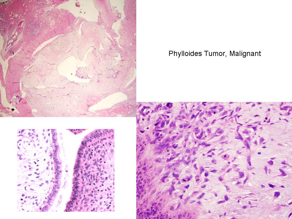
[Phylloides tumor]
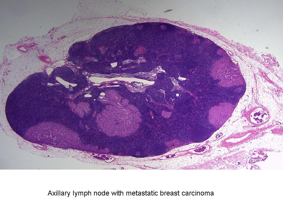
[Metastatic carcinoma]
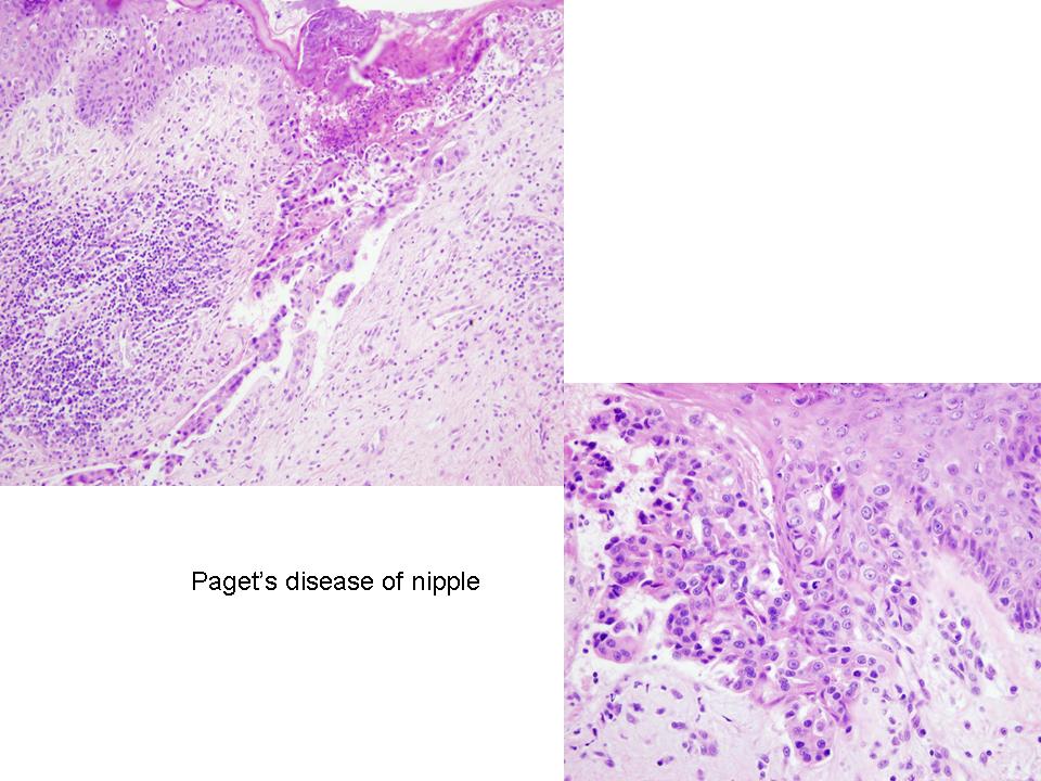
[Paget's disease]


