


Virtual Microscopy
|
|
||||
|
Virtual Microscopy |
||||
Case 1:
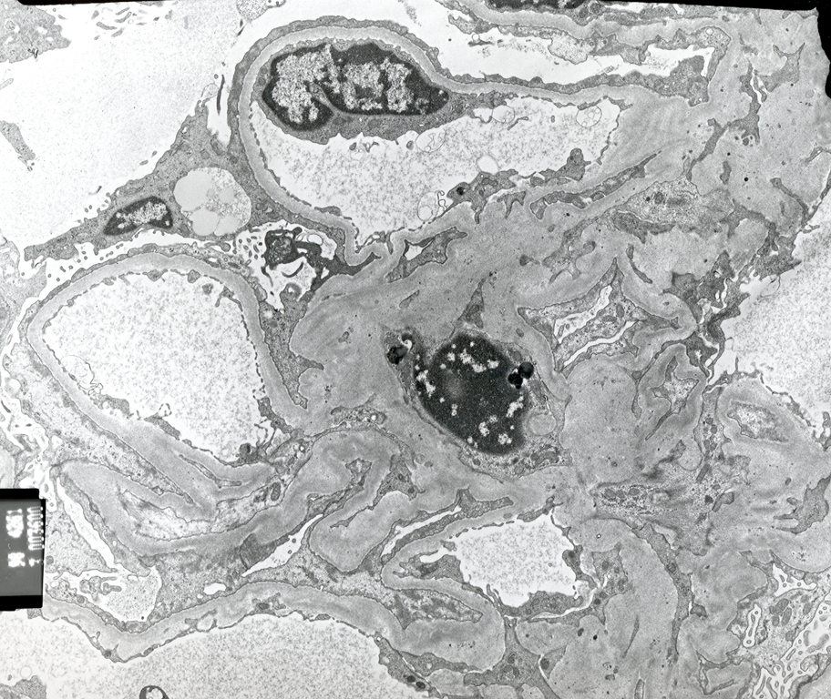
An 18-year-old male was referred for evaluation of proteinuria. He was trying out for the freshman college football team and the proteinuria was discovered during a routine physical examination. The proteinuria was subsequently quantitated to 3.0g/24hr and he was referred for further evaluation. During history and physical examination, he stated that he has noticed swelling of his lower extremity and has not been feeling as well as usual. A kidney biopsy was performed and it was interpreted as representing minimal change disease. Your slide is a representation of normal kidney. One electron micrograph is from a normal glomerulus and the other is from a patient with minimal change disease.
Slides: [normal kidney] [minimal change disease]
Electron micrographs: [normal glomerulus] [minimal change disease]
Case 2:
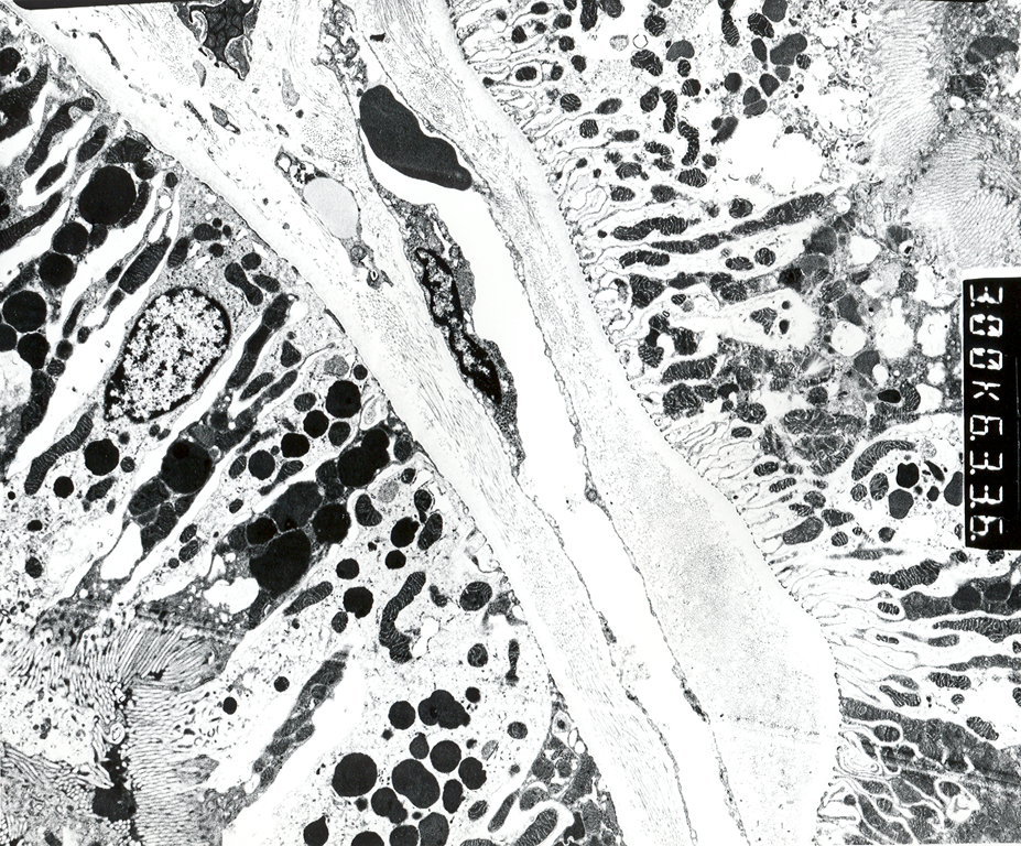
A 75-year-old female who has many medical problems developed flu like illness. She had diarrhea and complained of loss of appetite. She developed acute renal failure and a diagnosis of acute tubular necrosis was made. The histologic findings in acute tubular necrosis may be limited, but it allows us the opportunity to review renal tubular histology.
Electron Micrograph: [normal renal tubule]
Case 3:
A 45-year-old male presents with right sided flank pain. He has normal urine output and no significant prior health problems. His pain was only partially relieved by narcotic analgesics. He came to the emergency room and a small calculus was identified in his ureter. Four hours later the stone was passed in his urine and sent for analysis. He had an uneventful recovery. Review your histologic section of ureter.
Slide: [normal ureter]
.jpg)