


Virtual Microscopy
|
|
||||
|
Virtual Microscopy |
||||
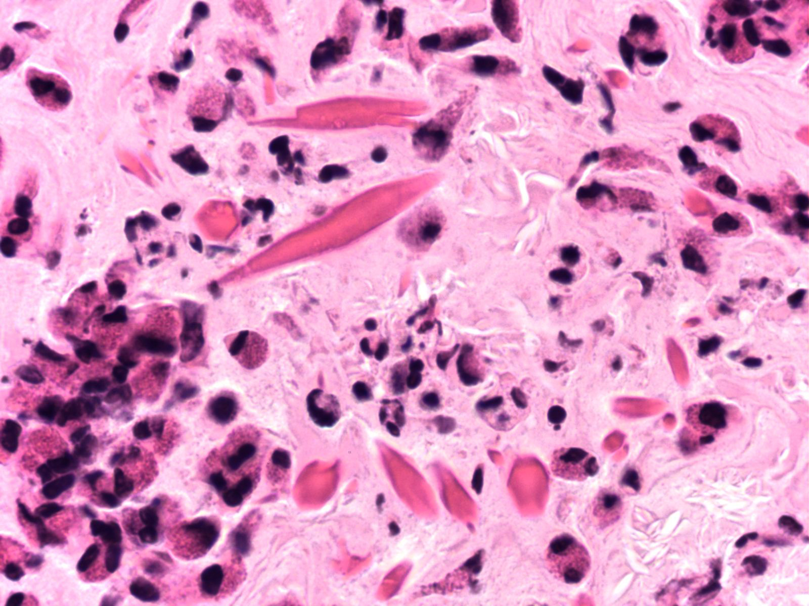
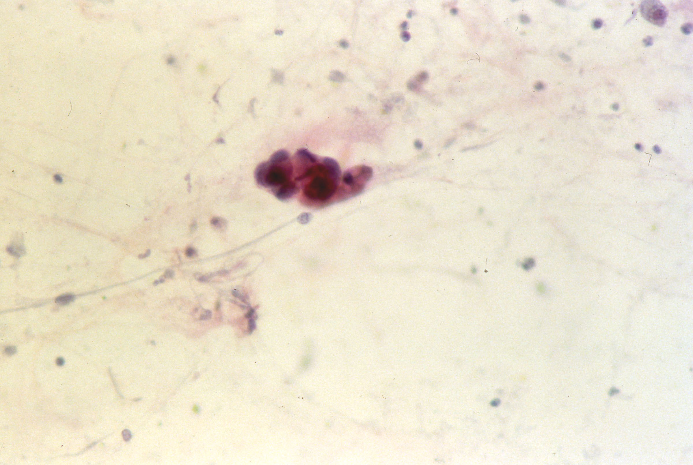
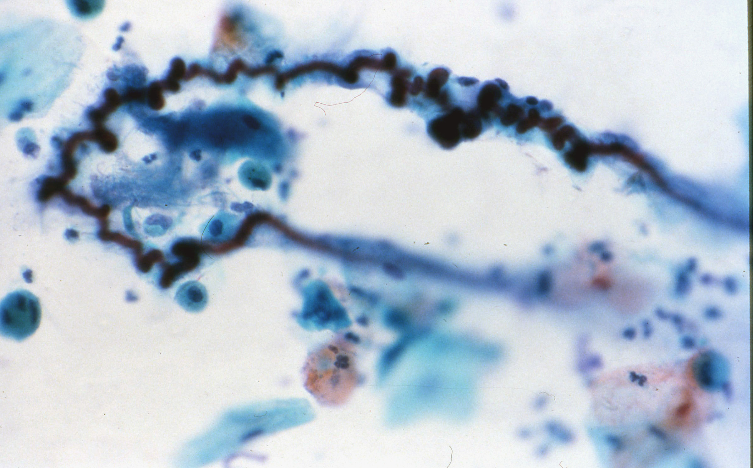
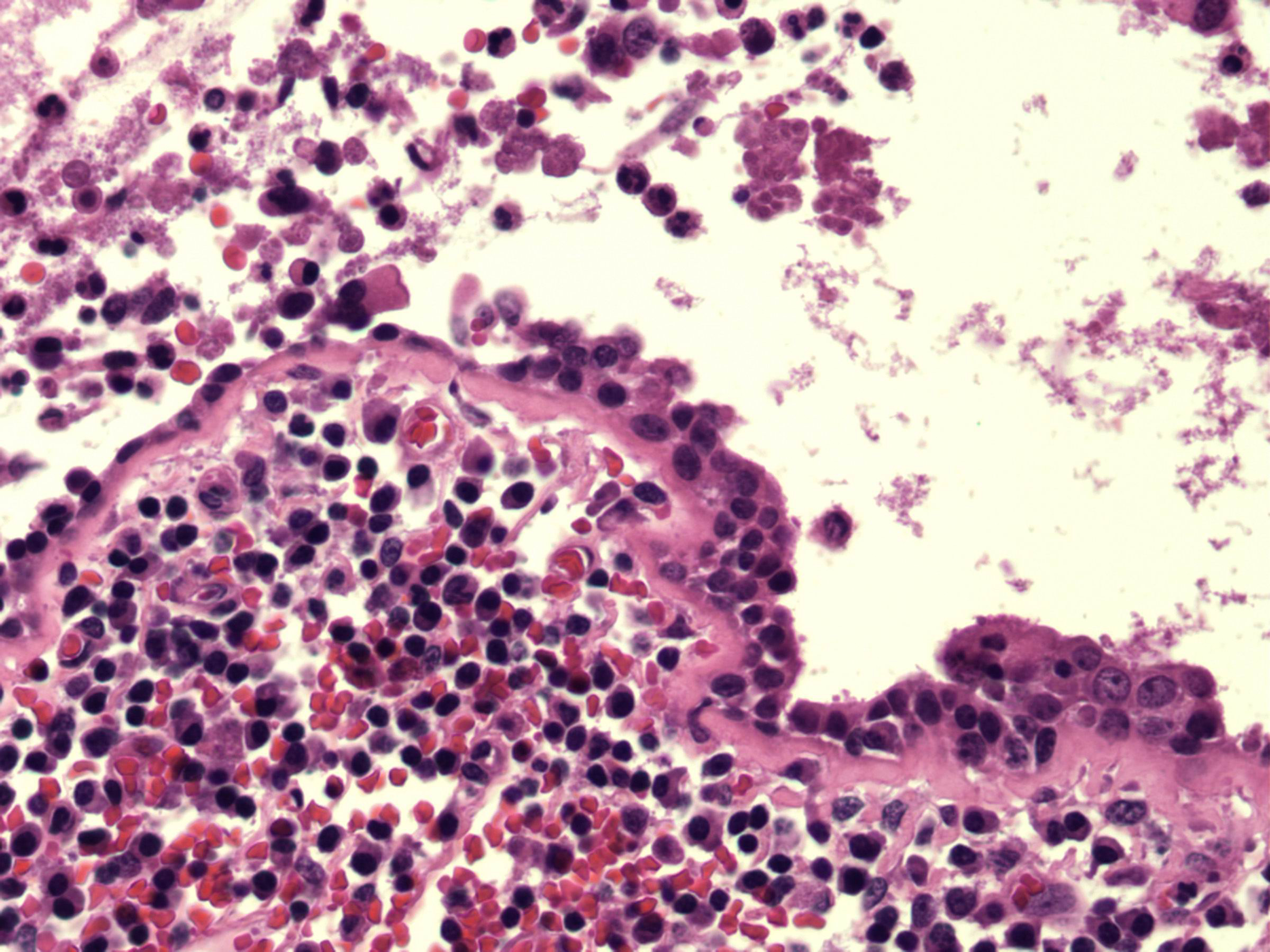
Case 1:
A 35 year old African American female presents with 10 year history of wheezing and shortness of breath with exercise.
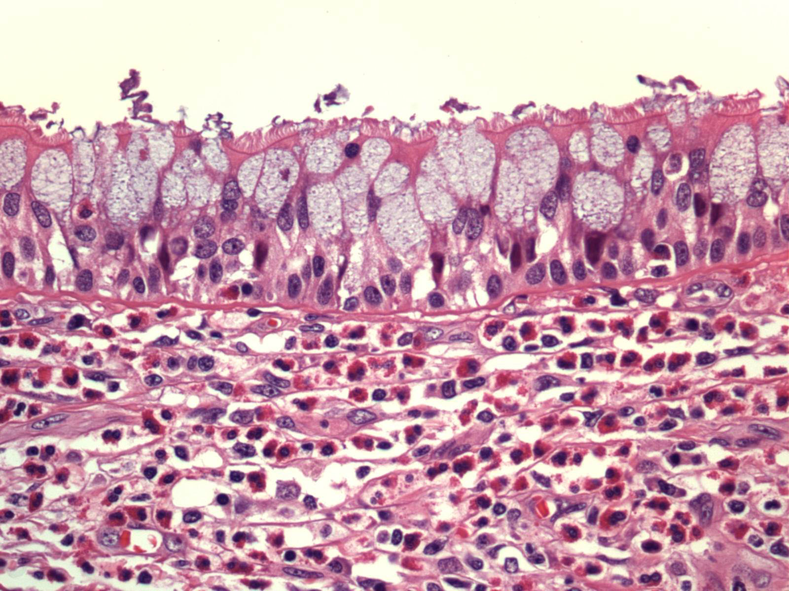
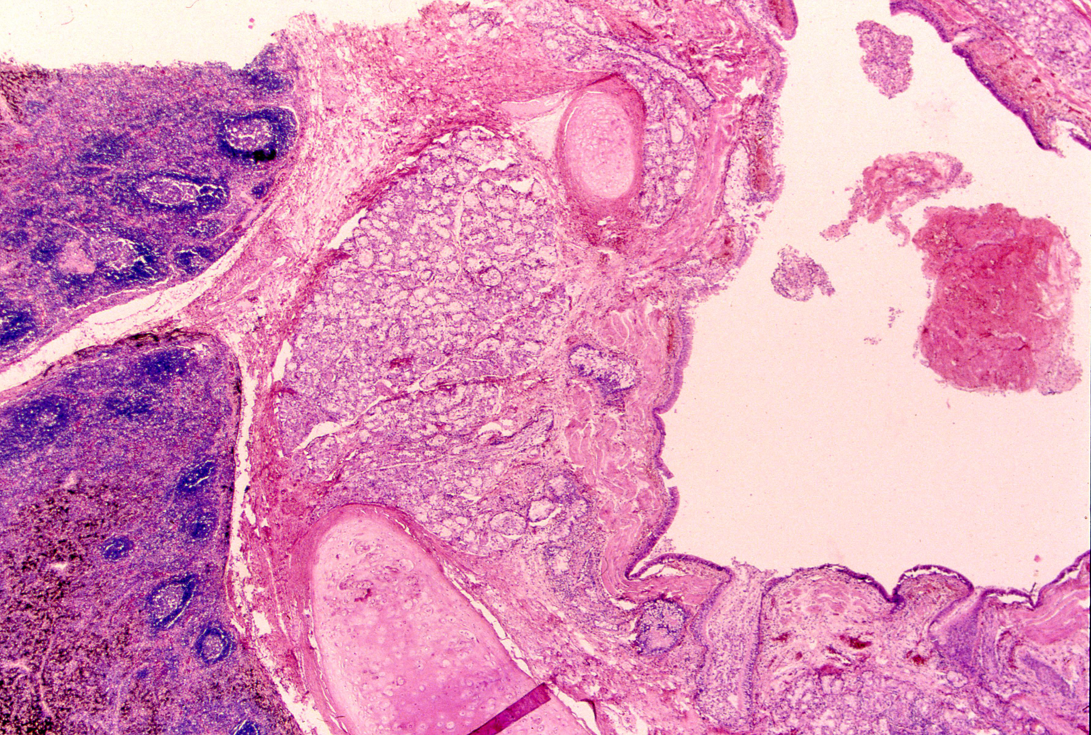
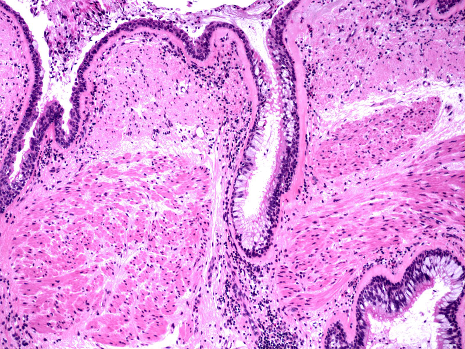
Slides: [Allergis mucin with Charcot-Leyden crystals] [Creola bodies] [Curshman's spiral] [Inflamed airway] [Large airway mucosa with goblet cell hyperplasia and eosino] [Low magnification asthmatic airway] [Mucous plug in proximal airway of asthmatic patient] [Thickened basement membrane]
Case 2:
A 18 year old Caucasian male presents with chronic pneumonias since childhood.
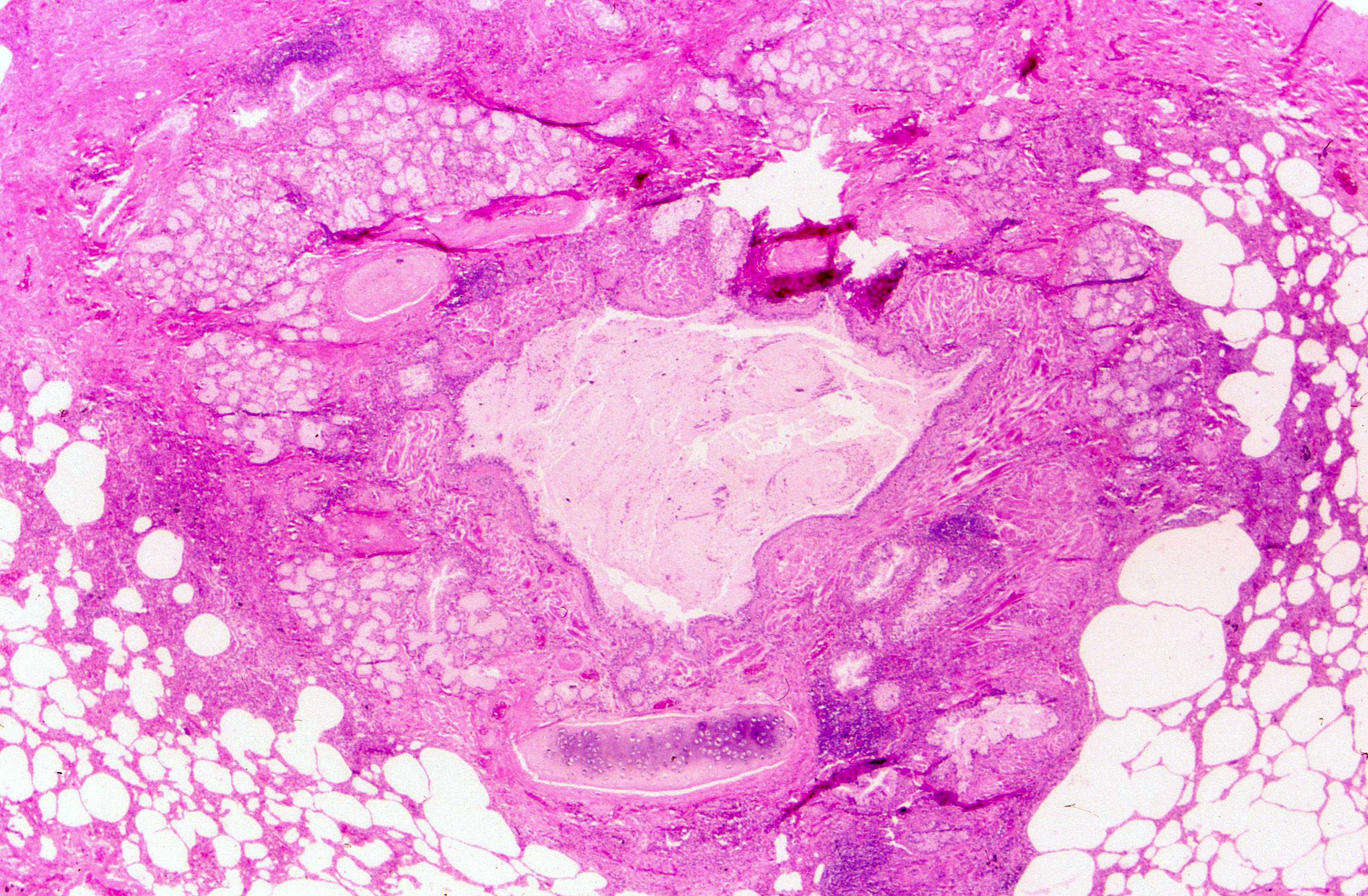
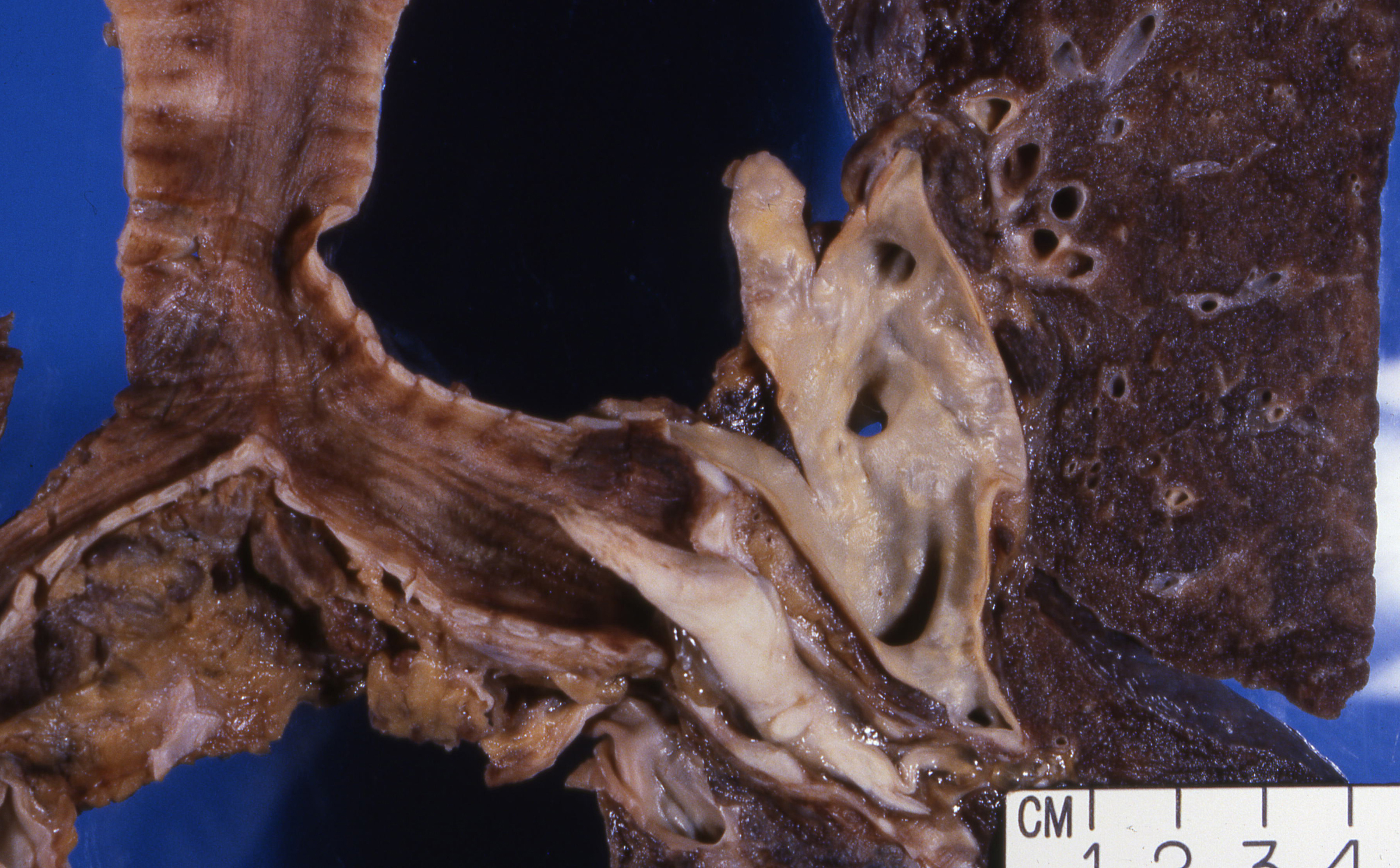
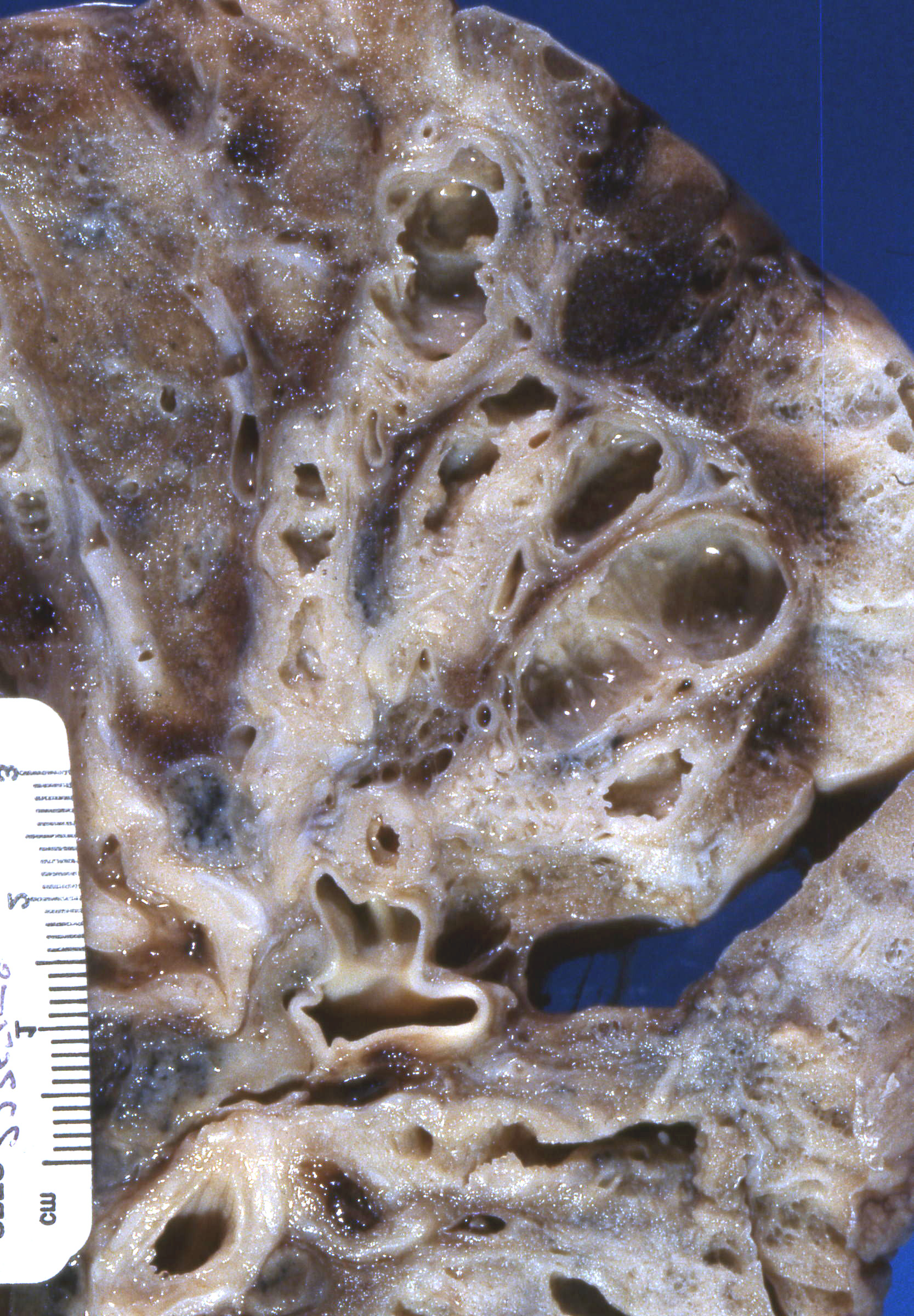
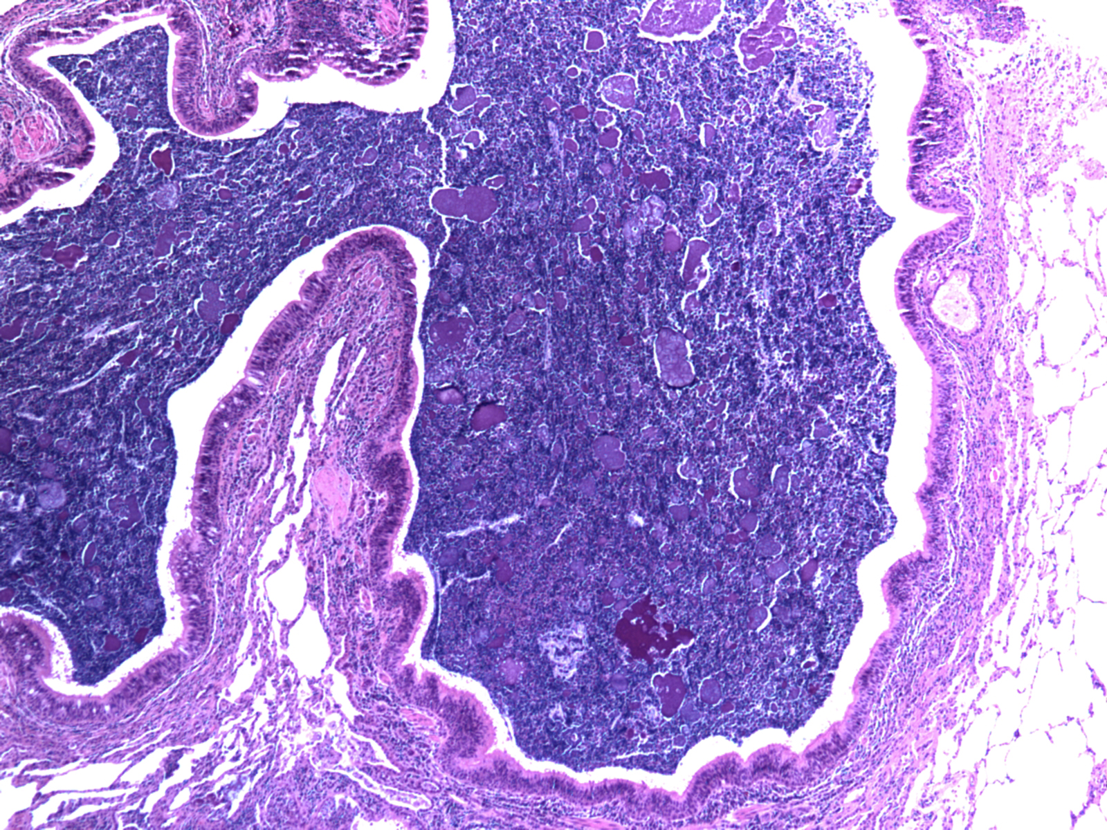
Slides: [Bronchiectasis (gross)] [Bronchiectatic airway with mucous plug] [Cystic fibrosis (gross)] [Diffuse bronchiectasis]
Case 3:
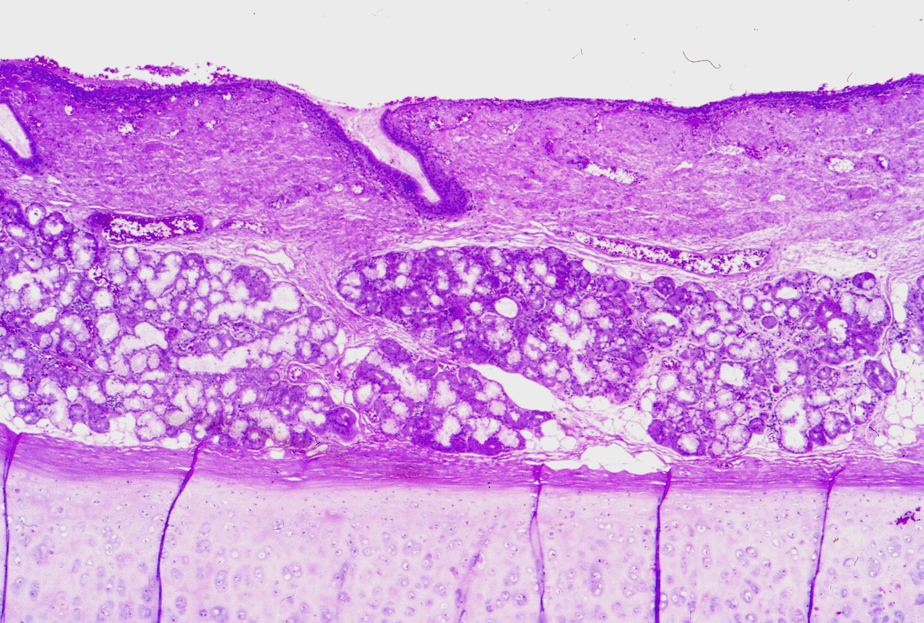
A 62 year old Caucasian male presents with 40-pack-a-year history of smoking.
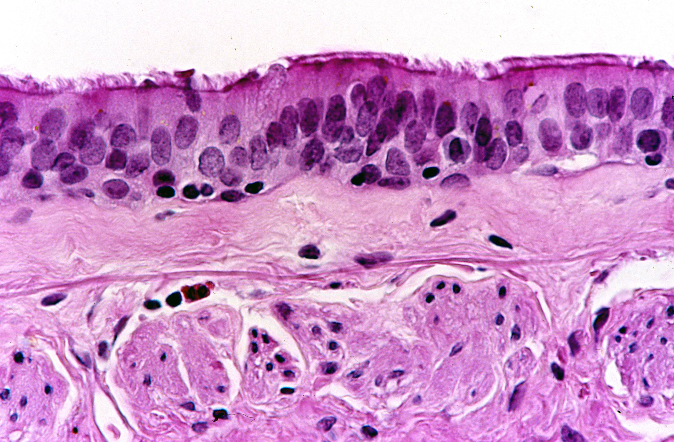
Slides: [Submucosal gland hyperplasia] [Submucosal gland hyperplasia (measure RI)] [Chronic bronchitis (smooth muscle hypertrophy)]
Case 4:
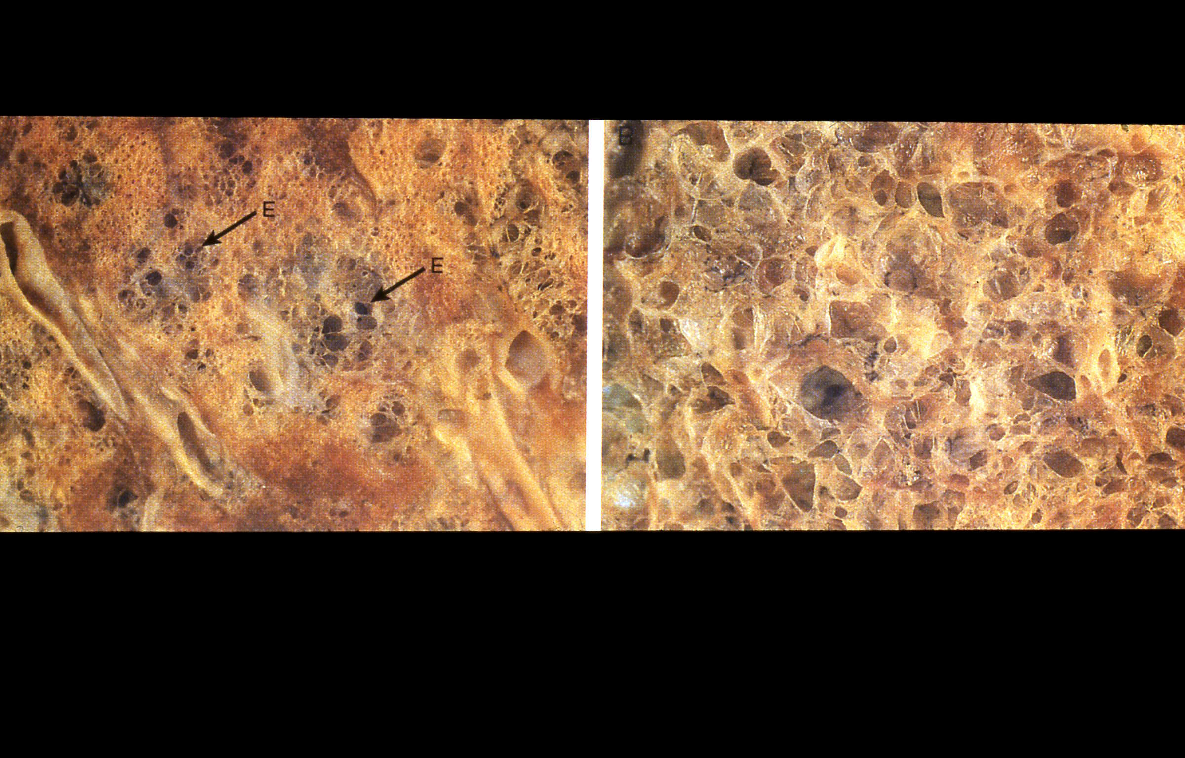
A 78 year old Caucasian female presents with 30-pack-a-year history and shortness of breath with exertion.
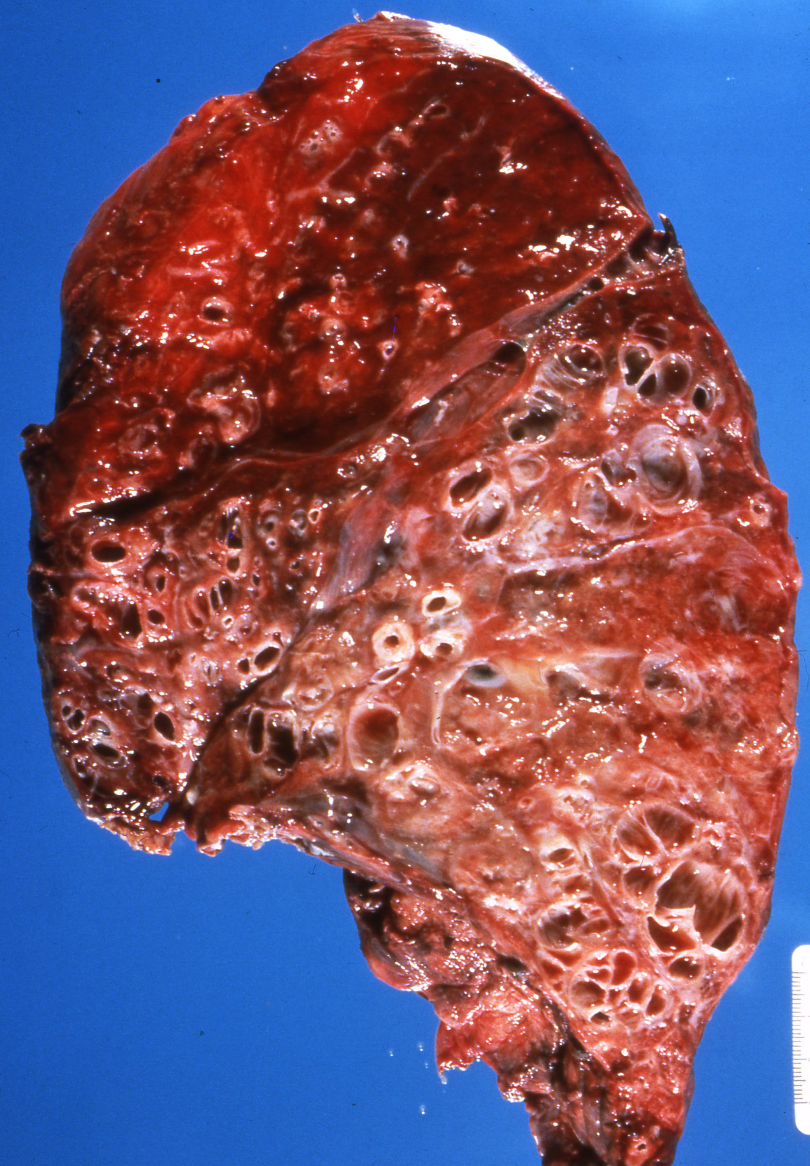
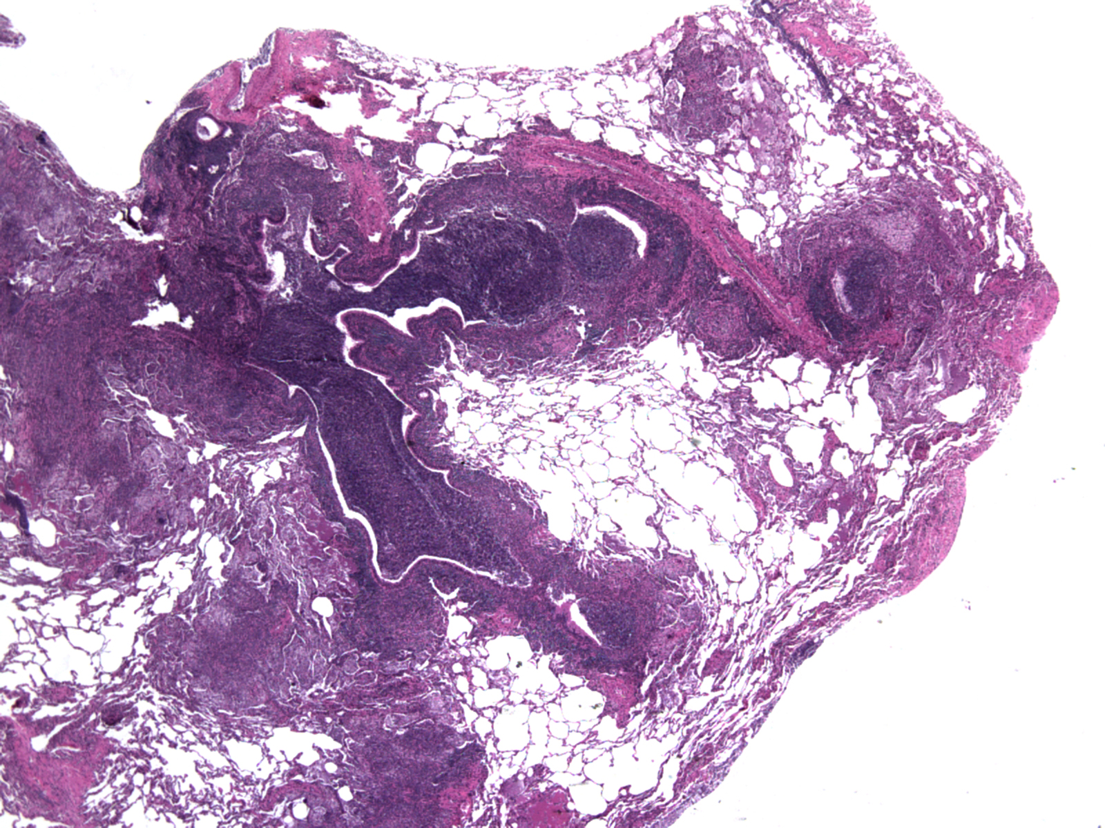
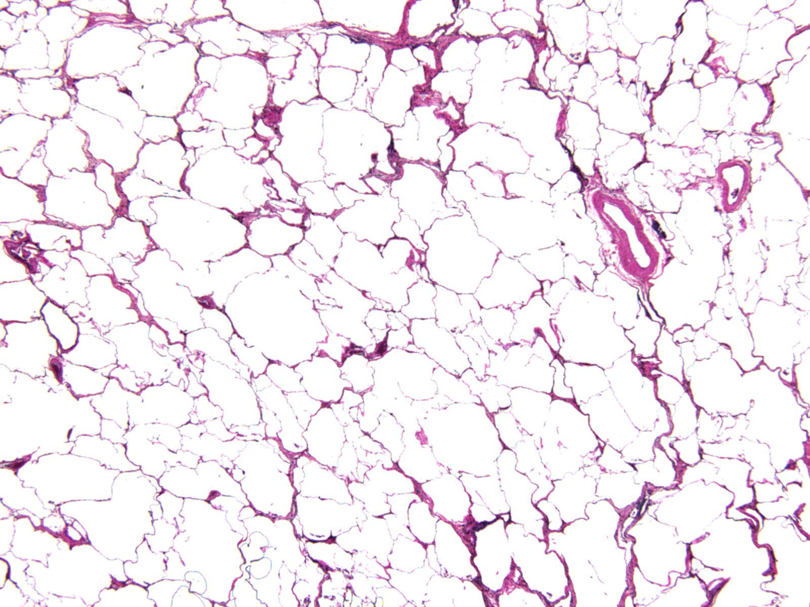
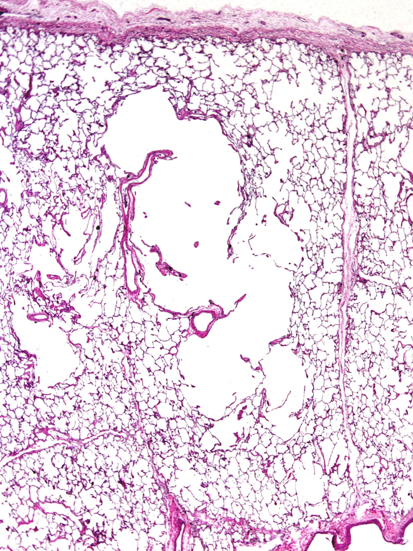
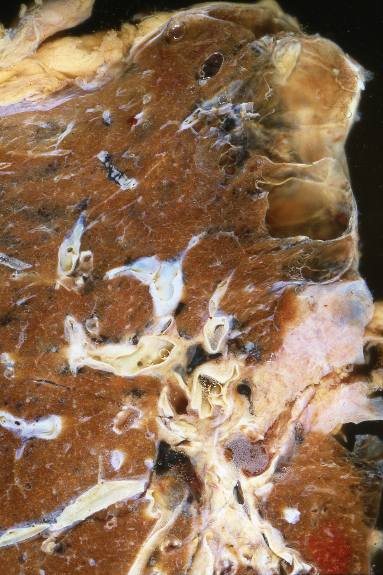
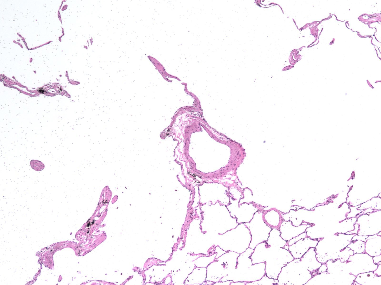
Slides: [Centriacinar and panacrinar emphysema] [Panacinar emphysema] [Centrilobular emphysema with lobule (low magnification)] [Emphysema bulla (gross)] [Pigmented macrophages of emphysema] [Loss of elastic recoil]