


Virtual Microscopy
|
|
||||
|
Virtual Microscopy |
||||
Case 1:
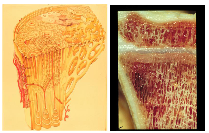
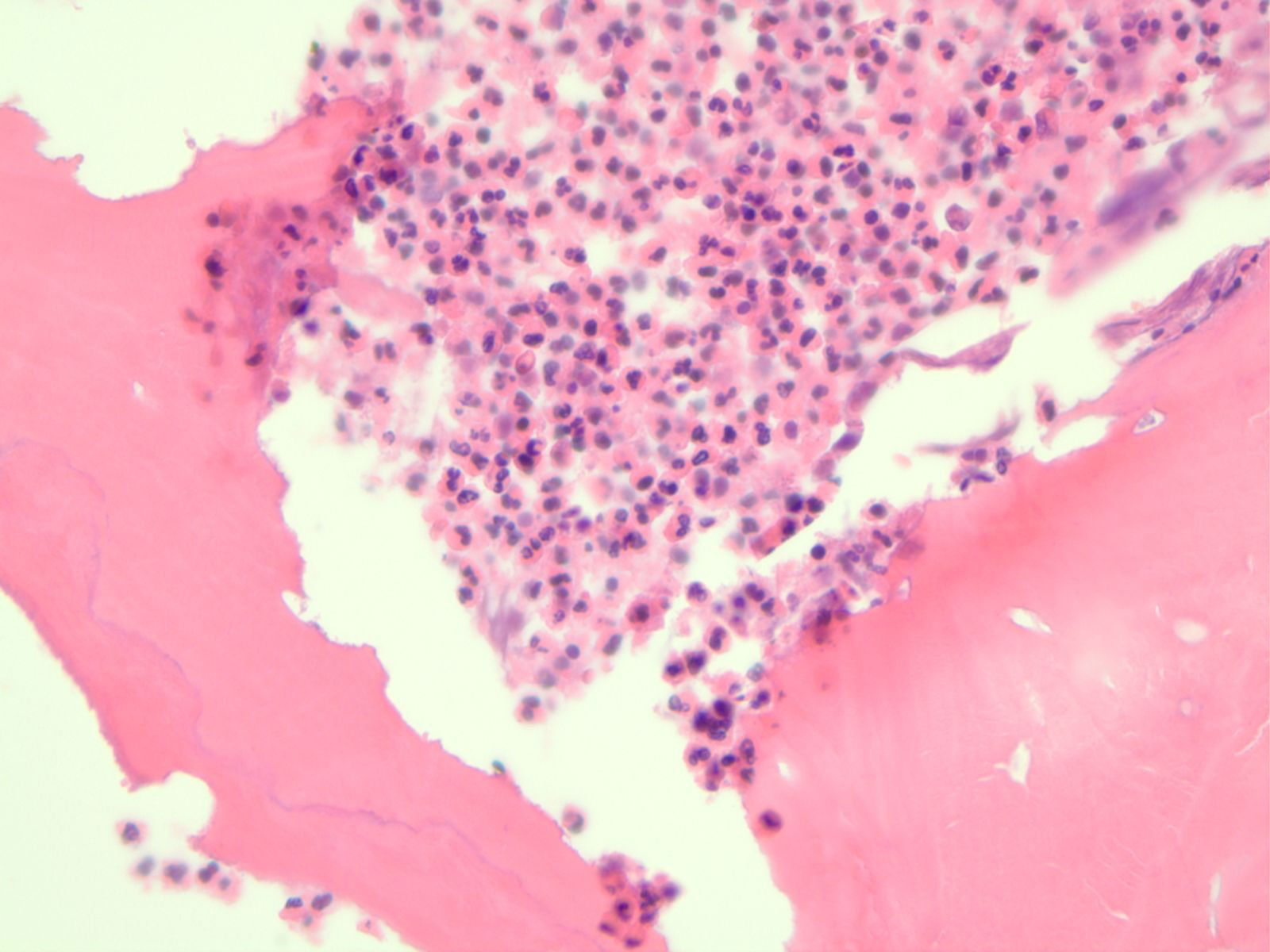
A 12 year old boy with a recent “cold” complains of left leg pain. The skin in the region of the pain is red and warm to the touch. The child has an elevated white cell count and an elevated erythrocyte sedimentation rate. Radiographic analysis revealed a sclerotic lesion involving the bony cortex.
Slide: [schematic of normal bone]
Photomicrograph: [acute osteomyelitis]
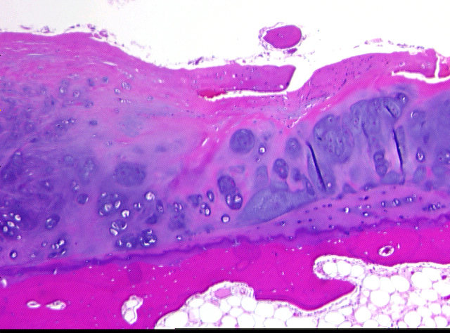
Case 2:
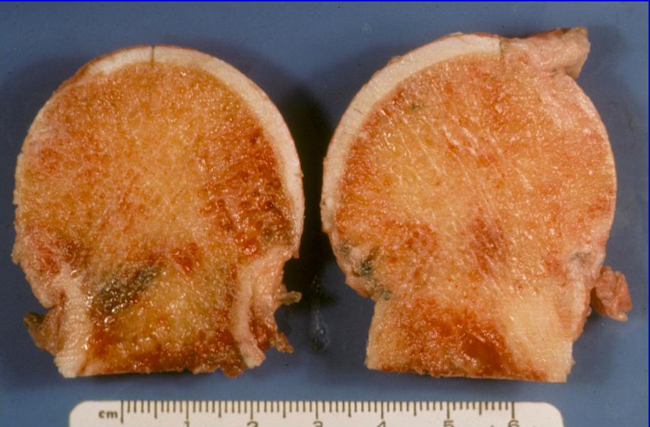
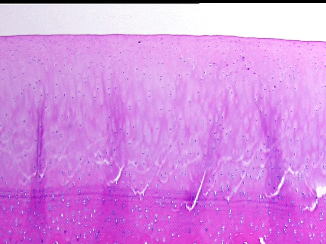
A 57-year old ex-NFL quarterback has had progressive pain and loss of movement in his hips bilaterally. Radiographic analysis revealed asymmetric joint space narrowing, subchondral sclerosis, and osteophyte formation.
Gross Photo: [gross picture of hip bones]
Photomicrograph: [normal articular surface]
Photomicrograph: [hip with loss of articular cartilage/eburnation]
Photomicrograph: [rheumatoid arthritis 1 and rheumatoid arthritis 2]
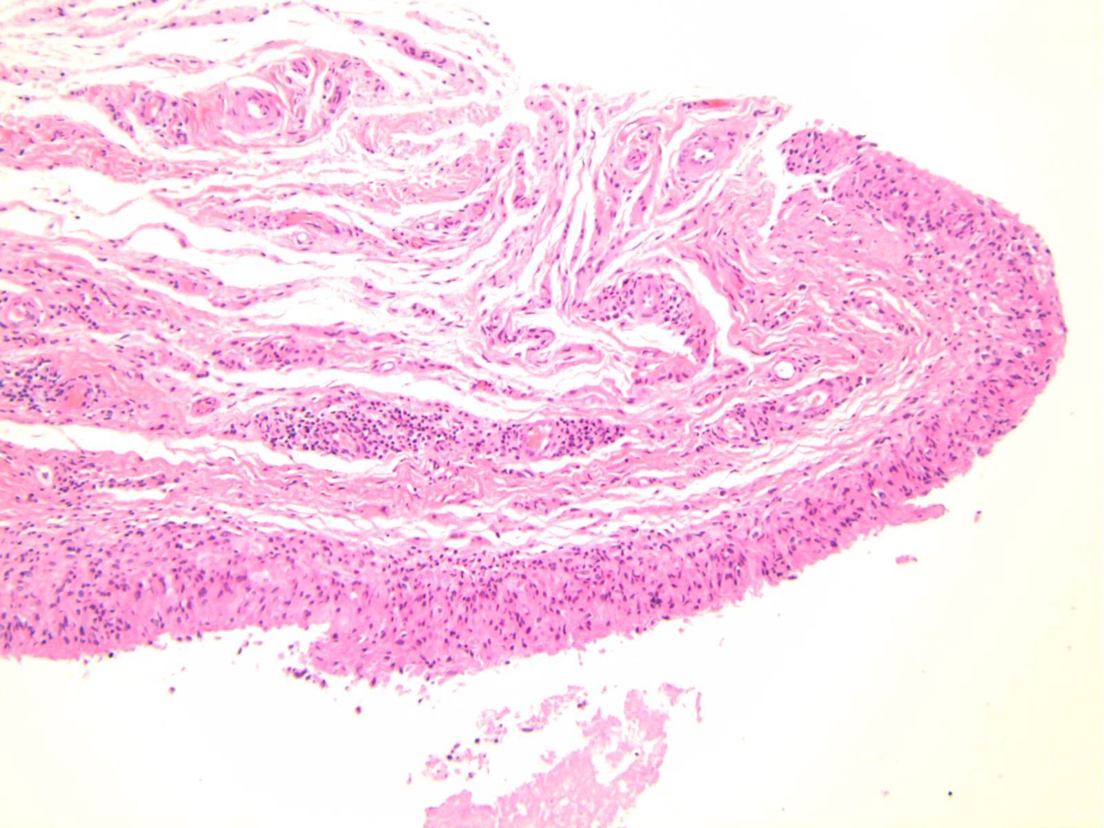
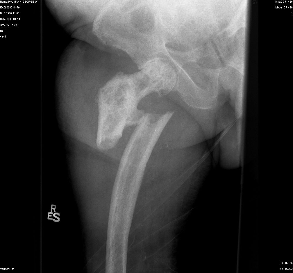
Case 3:
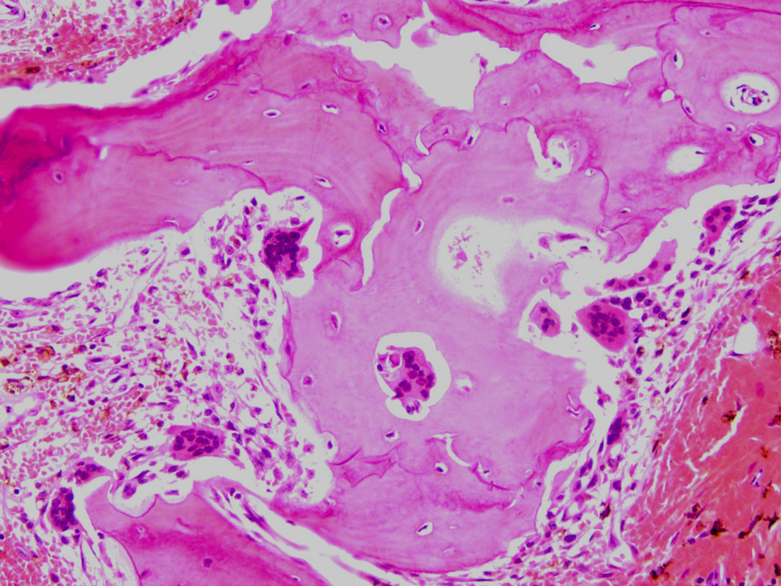
A 70 year old man with long standing chronic pain involving the right hip that has not been relieved by anti-pain medications, presents to the ER with a fracture of his right femur after stepping off a curb.
Slide: [radiograph of right femur]
Photomicrograph: [active Paget's disease]
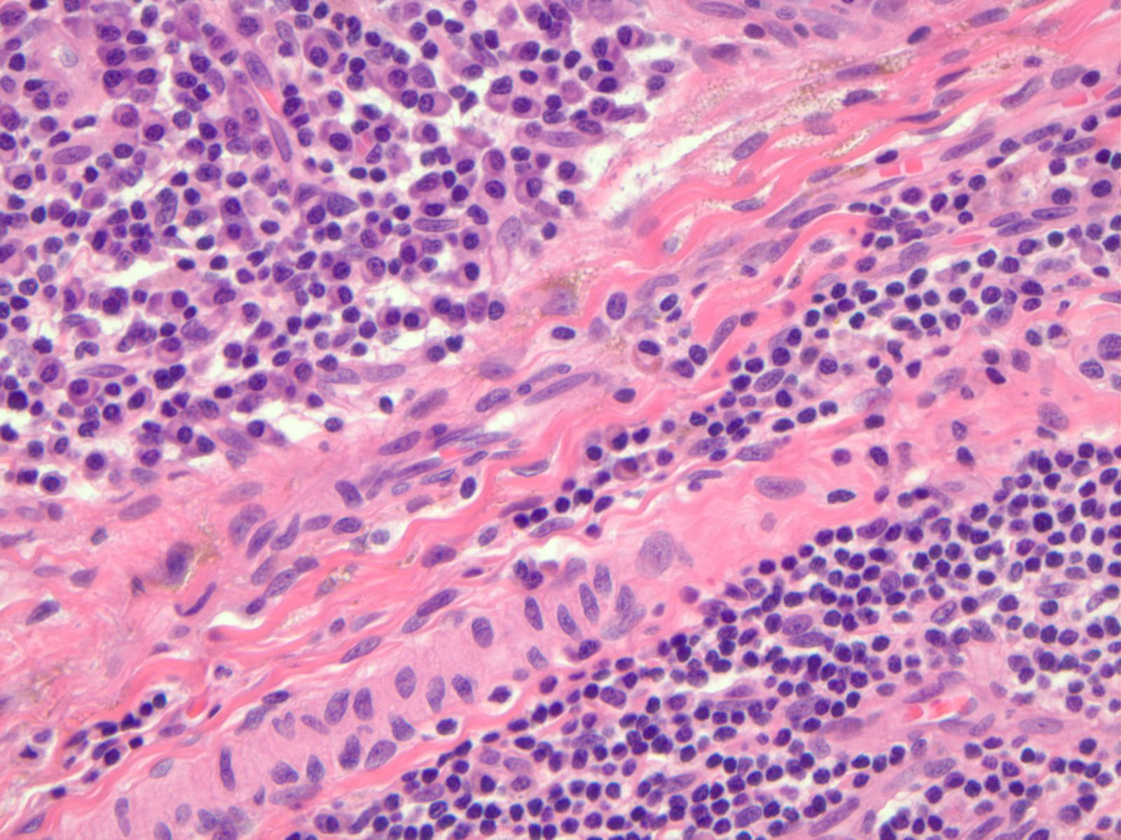
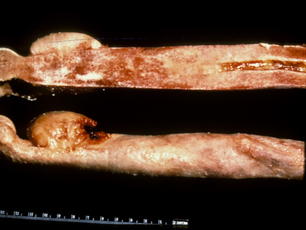
The patient is in for a routine checkup for management of his disease several years later and complains of severe pain in the right hip. After a workup, a major surery is performed to resect the femur.
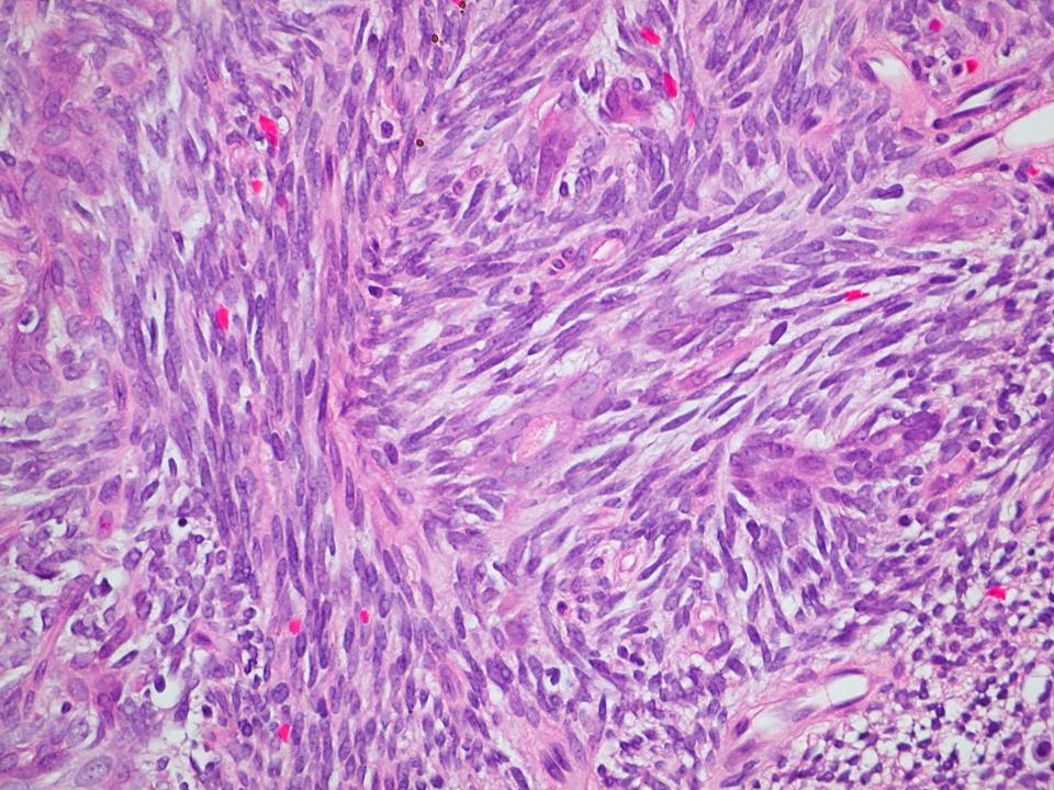
Gross Photo: [Paget's sarcoma]
Photomicrograph: [Paget's sarcoma]