Virtual Microscopy > Neuro > Brain Tumors
Case 1:
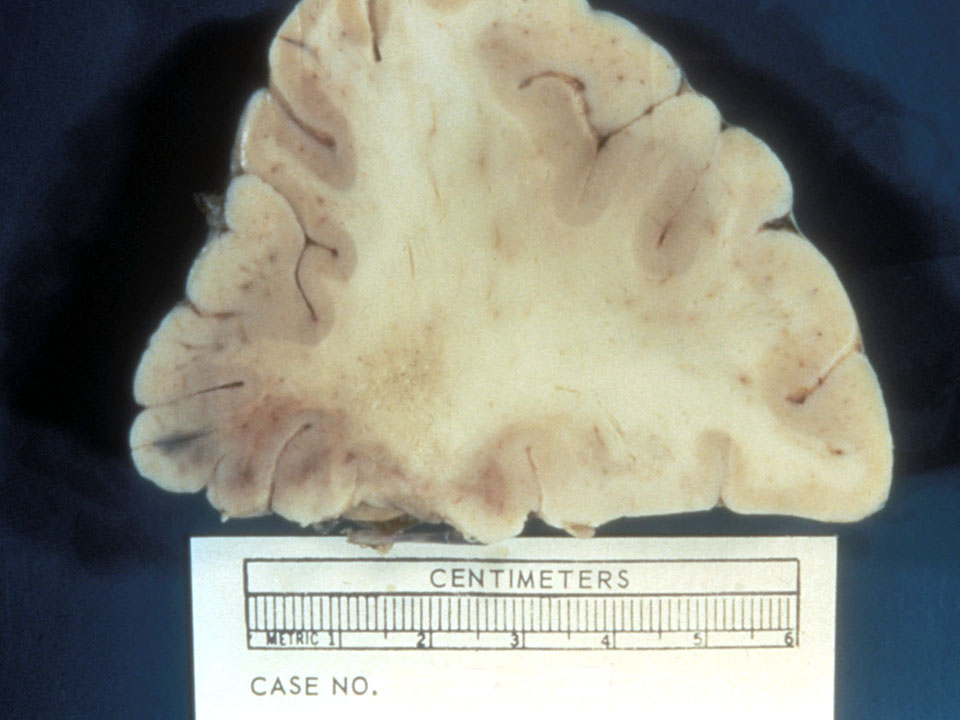
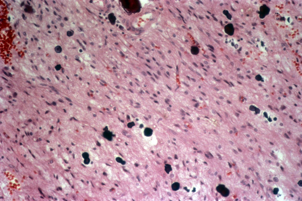
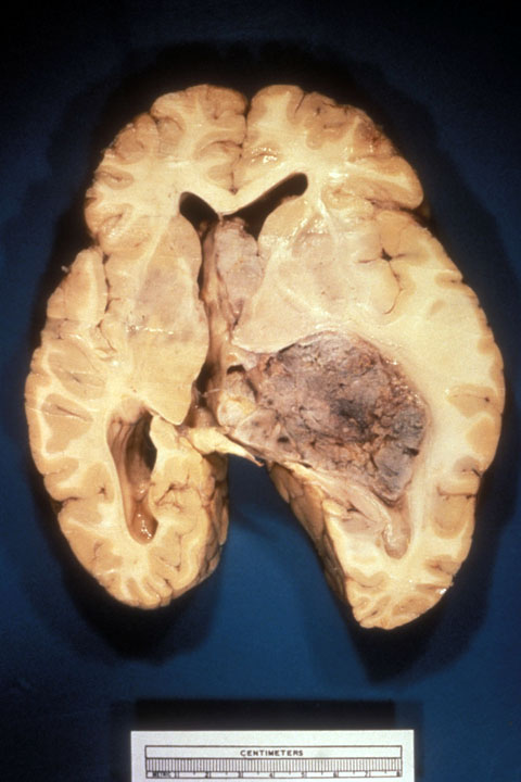
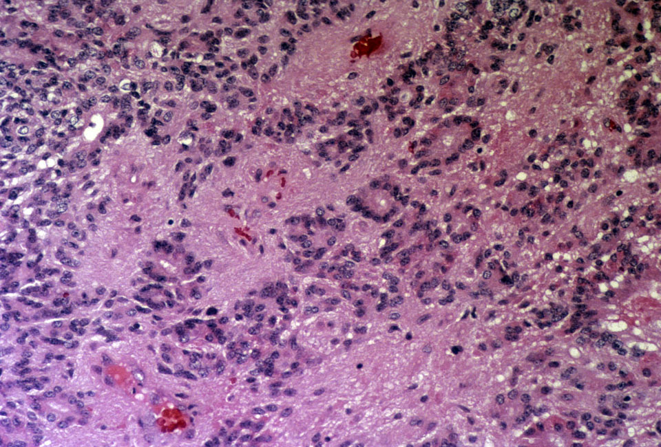
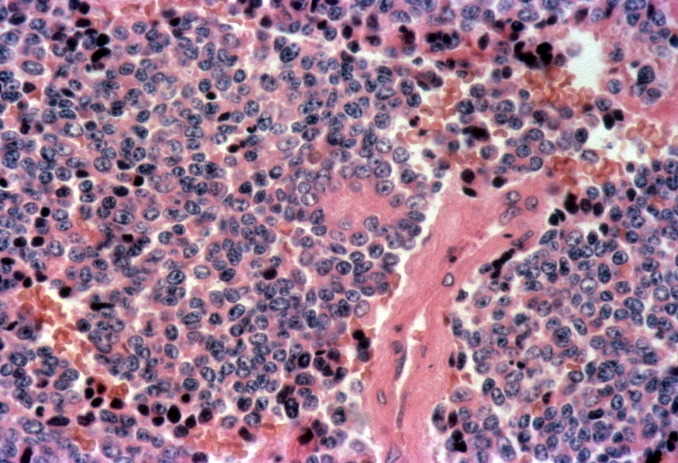
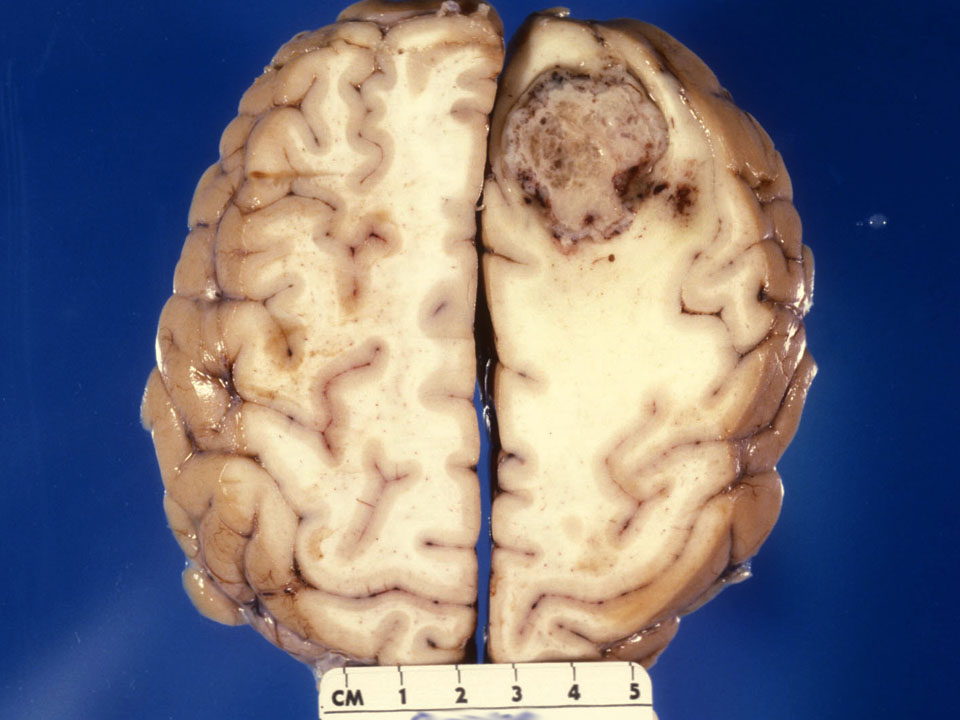
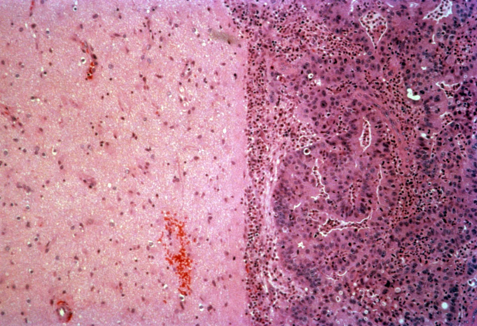
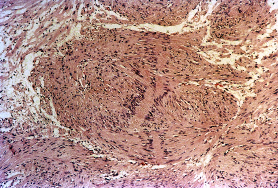
A 36-year-old male presented 6 years ago with a seizure and non-enhancing lesion in the frontal lobe. A subtotal resection of the lesion was performed. The gross and microscopic appearance of the lesion are shown here [gross patient's tumor] [microscopic patient's tumor]. The tumor more recently is increasing in size and shows ring-enhancement. A biopsy is taken [virtual slide of patient's tumor on biopsy]
- Describe the gross findings in the brain at the time of initial resection? (see gross of patient's tumor)
- How does the microscopic appearance of the original lesion compare with the recent appearance of the tumor? (compare microscopic of patient's tumor with virtual slide of patient's tumor on biopsy)
- What is the most likely cell of origin for this tumor? How might you confirm your answer?
Case 2:
The patient in case 1 has a 28-year-old sister who presents with seizures and a fronto-parietal lobe mass. A biopsy is taken.
Slide: [virtual slide of patient]
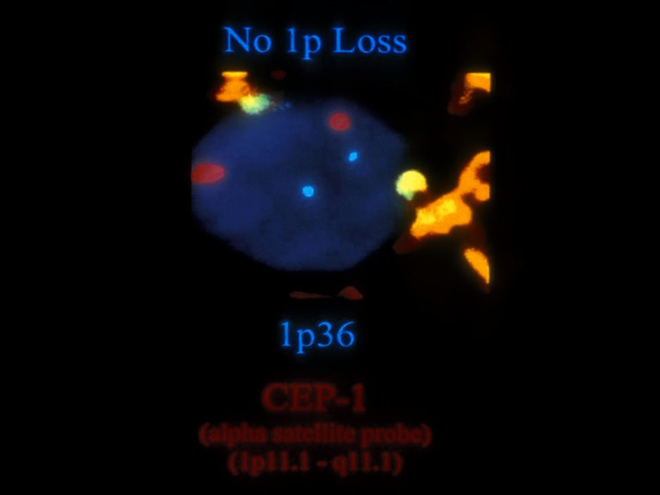
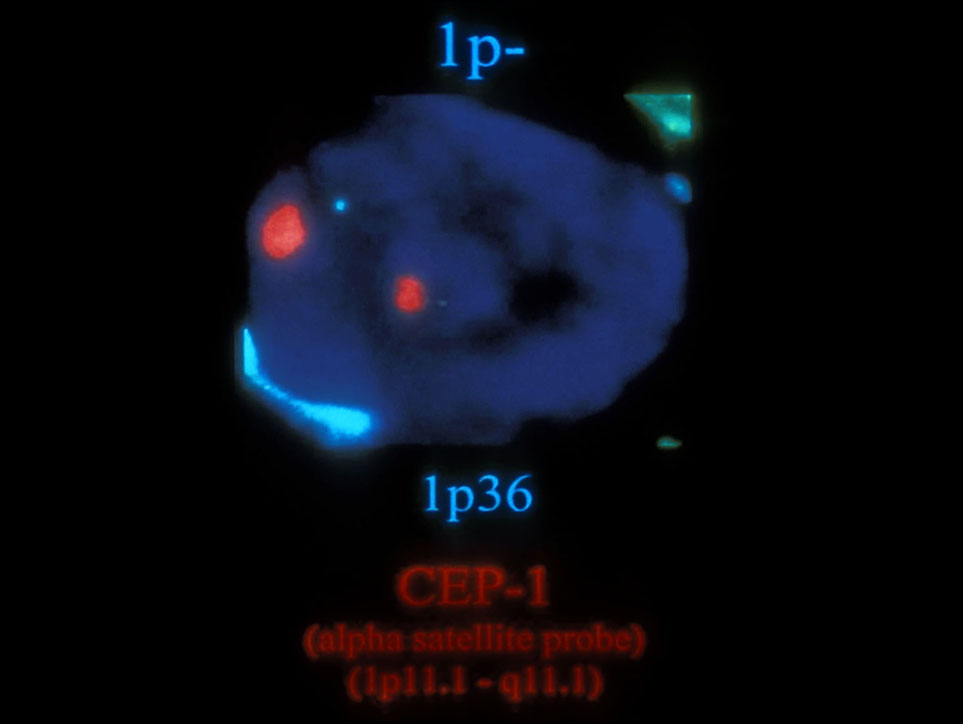
Photomicrographs: [1p normal FISH] [1p deleted FISH]
- Describe the morphologic features of this tumor? How is this the same or different than the tumor in case 1.
- What is the most likely cell of origin? Where would you guess is the most likely location for this tumor to arise?
- What is the clinical significance of distinguishing this tumor from the tumor in case 1.
Case 3:
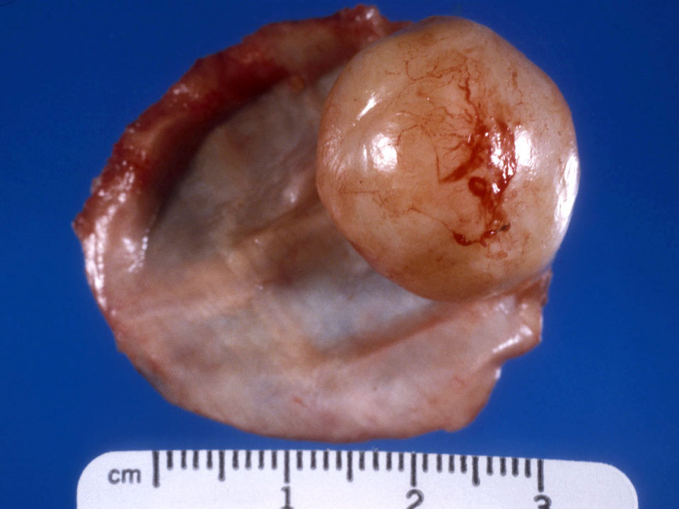
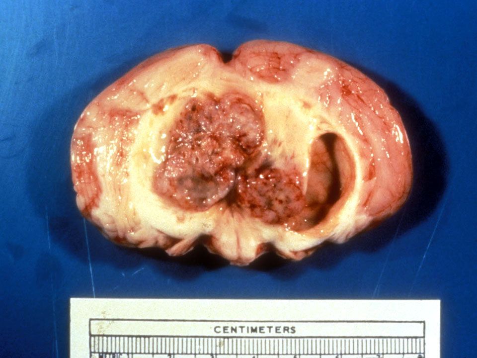
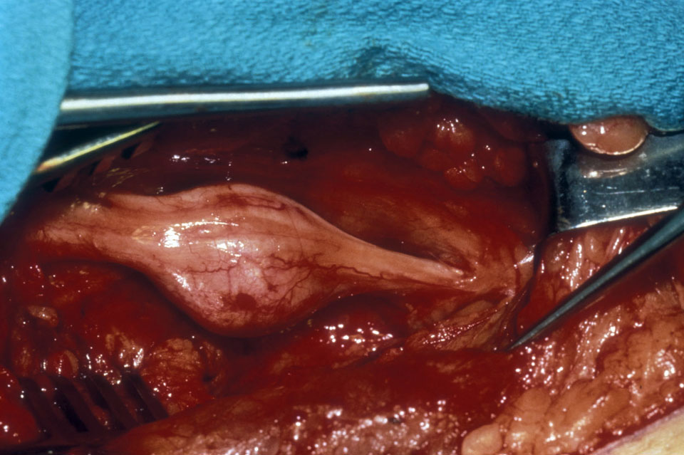
The patient in case 1 has a 67-year-old grandmother who presents with recurrent headaches. On imaging, she has dural-based mass. The tumor is completely excised.
Gross photograph: [patient's tumor]
Slide: [virtual slide of patient's tumor]
- Describe the gross appearance of the tumor?
- Given the location of the tumor, what is the most likely cell of origin?
- Given the histologic appearance of the tumor, would you expect it to behave more like a benign or malignant neoplasm? Why?
Show and Tell Cases:
- [ependymoma gross] [ependymoma microscopic]
- [medulloblastoma gross] [medulloblastoma micro]
- [metastasis gross] [metastasis micro]
- [schwannoma gross] [schwannoma micro]


