


Virtual Microscopy
|
|
||||
|
Virtual Microscopy |
||||
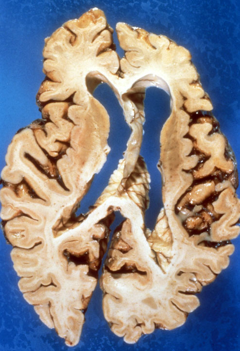
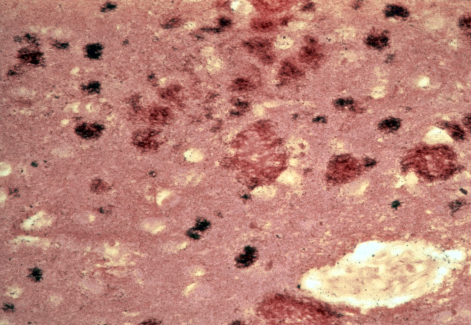
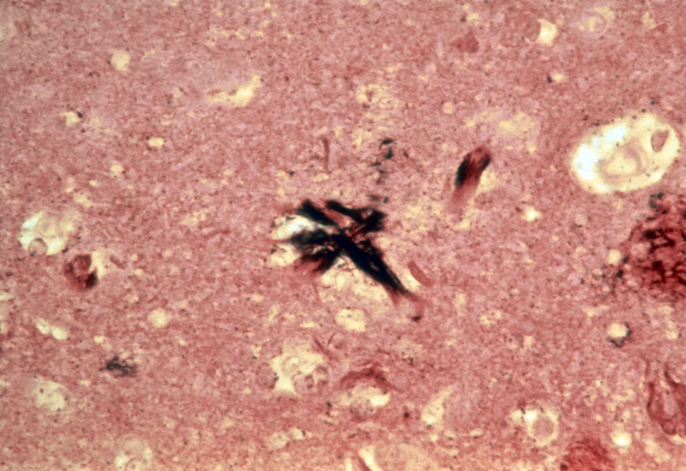
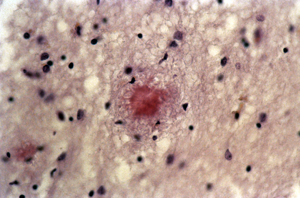
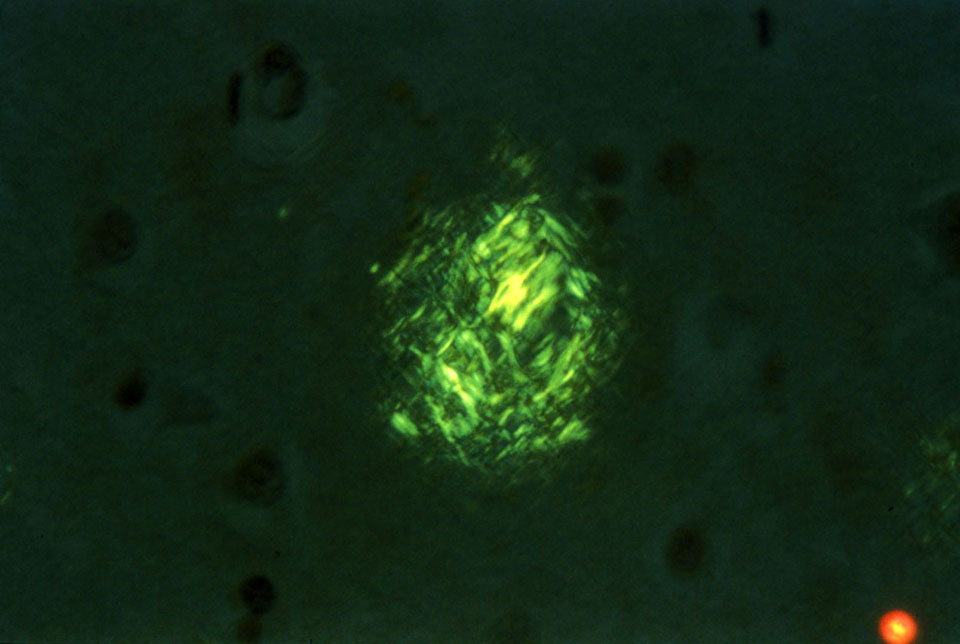
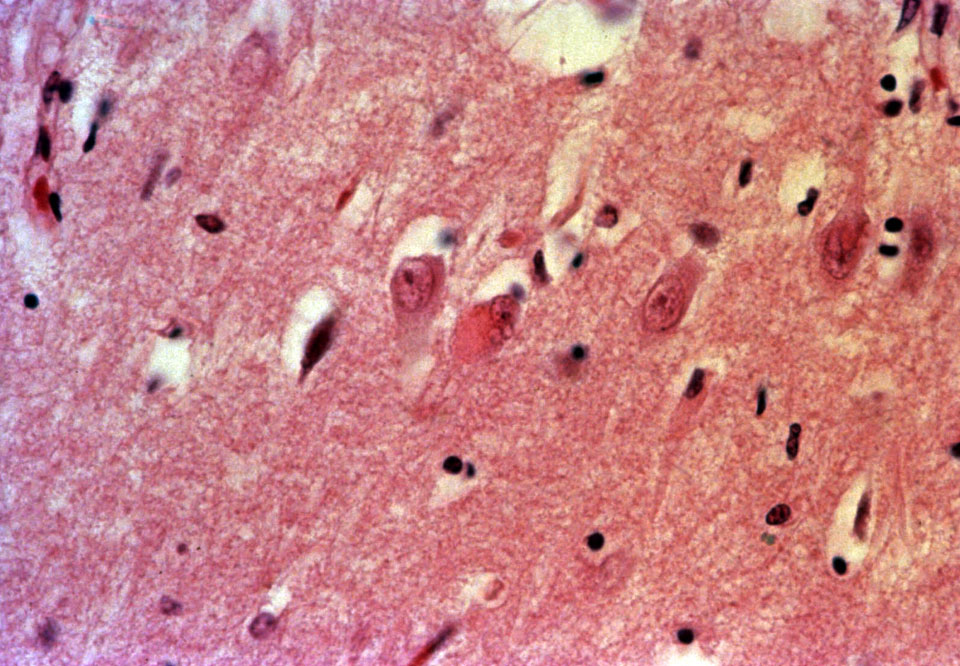
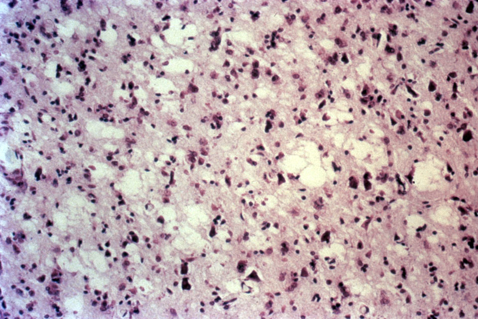
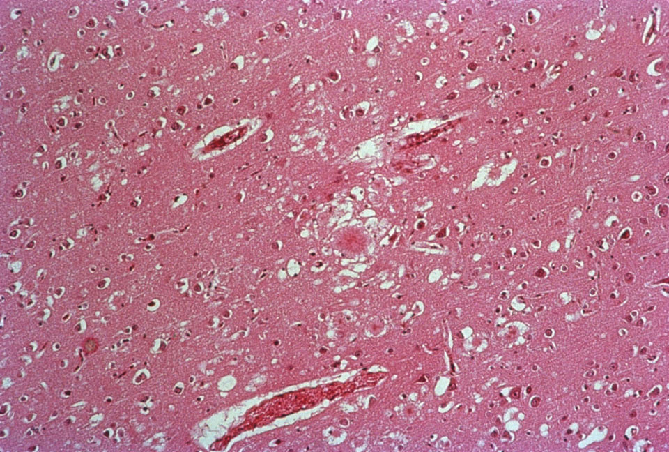
Case 1:
The patient is a 76-year-old female with a slowly progressive history of memory loss and dementia. She spent the last year of the life in a nursing home, requiring full nursing care.
Slides: [patient's brain (H&E stain)] [patient's brain (Bielschowsky stain)]
Gross photograph: [patient's brain]
Photomicrographs: [plaque (silver stain)] [tangle (silver stain)] [amyloid in plaque (congo red stain)] [amyloid in plaque (congo red stain, polarized] [granulovacuolar degeneration] [Hirano body]
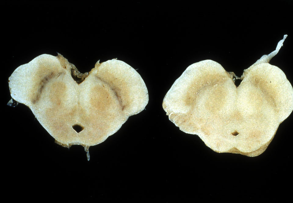
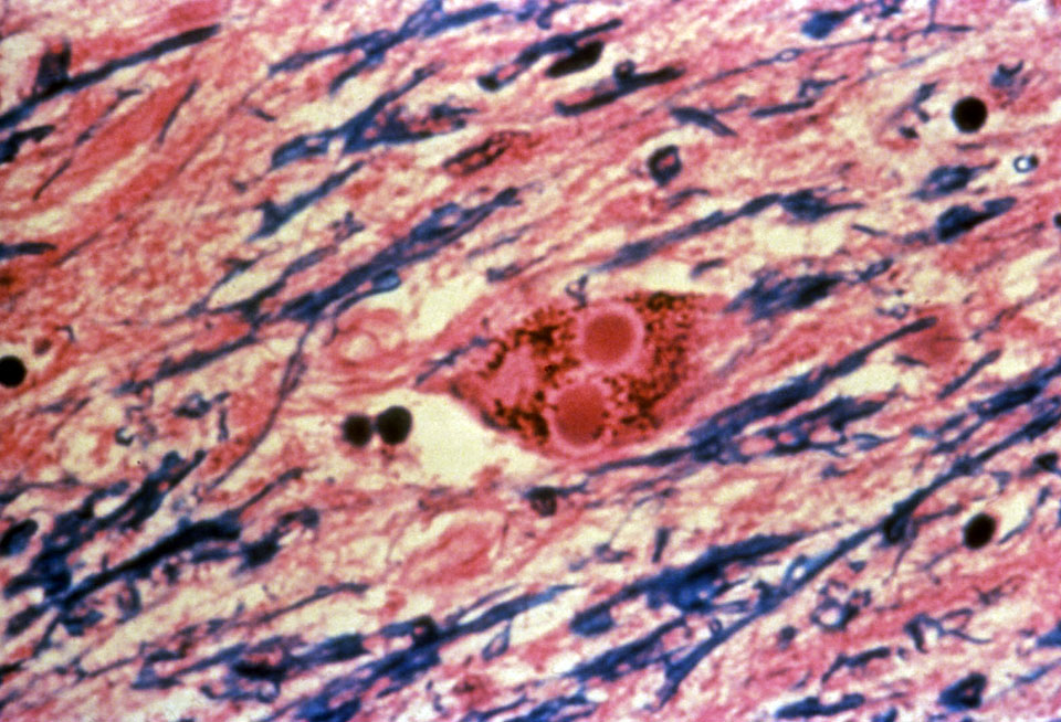
Case 2:
A 63-year-old male had a history of a pill-rolling tremor, unsteady gait, stooped posture and masked facies.
Gross photograph: [normal brain (left), patient's brain (right)]
Slide: [patient's brain]
Photomicrograph: [Lewy body]
Show & Tell Case: