Virtual Microscopy > Neuro > CNS Histology Teaching Cases
Case 1:
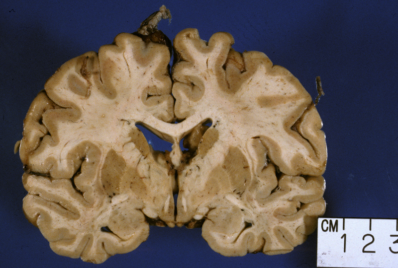
A 17-year-old male with normal development presented with a seizure. A
magnetic resonance imaging (MRI) study of the brain performed after the seizure
revealed the presence of thickening of the cortex in the right peri-insular
region.
Slides: [normal cerebral cortex (note the meninges are not normal in this slide)] [annotated cerebral cortex] [slide from this patient] [normal cerebral cortex]
Gross images: [normal gross image] [gross photograph of this specimen]
- Review the normal section of cerebral cortex (the meninges are not normal in this slide--there is meningitis). Identify meninges, gray matter (cortex), and white matter. What are the normal cellular constituents of each of these regions? Be able to identify arachnoid cap cells, neurons, astrocytes, oligodendrocytes, microglial cells, and blood vessels.
- Review the slide from this patient. Compare the normal cortex to the cortex in this patient. What differences do you see? Pay attention to the cortical layering, orientation of cells within the cortex and cell morphology.
- What might be the role or consequence of these alterations in terms of function/dysfunction of the cortex?
Case 2:
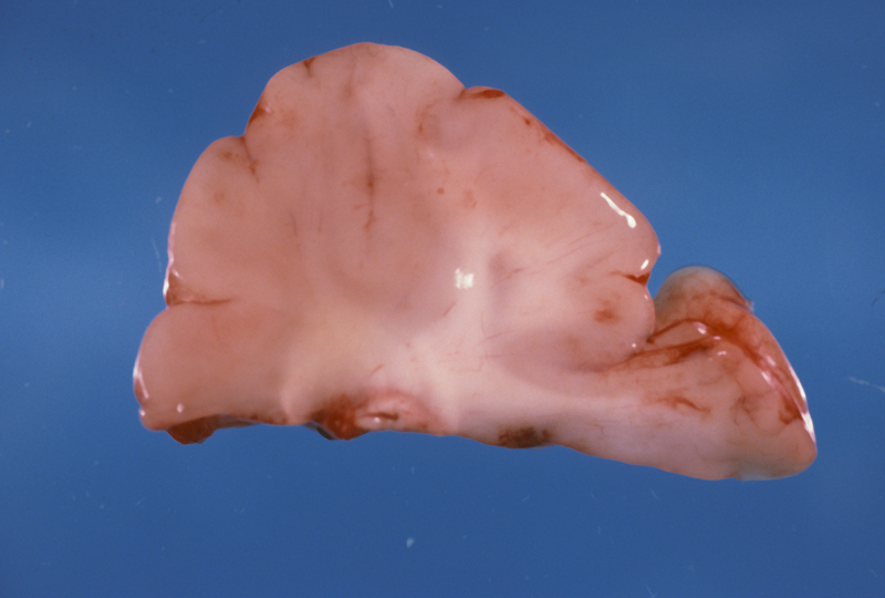
A 2-month old male born with a congenital heart disorder required surgery for
correction of his heart defect. After surgery, he became septic and died. A
section of cerebellum is evaluated at autopsy.
Slides: [Normal adult cerebellum] [annotated normal adult cerebellum] [this patient's cerebellum]
- Review slide of normal adult cerebellum. Identify the meninges, molecular layer, Purkinje cells,
granular cell layers, white matter, and dentate nucleus.
- Review slide of this patient's cerebellum. What is different about the histologic section of cerebellum from the
2-month old male?
- What is the significance of this finding, functionally and
developmentally?
Case 3:
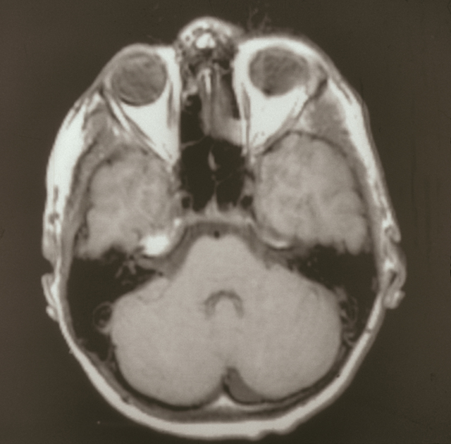
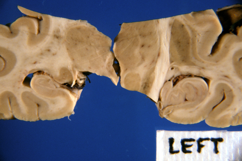
A 12-year old male presents with an 8 year history of seizures (epilepsy)
refractory to multiple different attempts at pharmacologic management. On
MRI studies, there is increased signal intensity in
the right hippocampal region and evidence of atrophy. To control the patient’s
seizures (which number between 12-15 a day), the patient undergoes a resection
of the right lateral temporal lobe, amygdala, and hippocampus. A histologic
section of the hippocampus is available for review.
Slides: [this patient's hippocampus] [annotated normal hippocampus] [normal section of hippocampus]
Gross and Radiographic images: [magnetic resonance imaging (MRI) image] [normal hippocampus]
- Review normal slide of hippocampus. Be able to identify the following normal structures in the hippocampal section: lateral ventricle, choroid plexus, dentate, endplate of hippocampus (CA4), CA3 sector, resistance sector (CA2), Sommer sector (CA1), and subiculum.
- There is a loss of neurons in the resected hippocampus in what regions? What cells proliferate in response to the loss of neurons?
- How might the loss of neurons result in seizures?
Case 4:
A 67-year old male presents with a 2 year history of progressive muscular
atrophy and limb weakness. Two months ago he develops difficulty swallowing (dysphagia).
Two weeks before his death, he requires mechanical ventilation (on a ventilator)
and dies of a pneumonia. An autopsy is performed and sections of his spinal cord
are examined.
Slides: [patient's spinal cord (stained with modified Kluver-Barrera, myelinated areas stain blue)] [annotated normal spinal cord] [normal slide of spinal cord] [normal spinal cord]
- Review normal section of spinal cord. Identify the following structures: meninges, gray matter, white matter, posterior columns, lateral
corticospinal tracts, ventral corticospinal tracts, central canal, anterior horn cells, anterior and posterior nerve roots.
- What cells line the central canal?
- What morphologic features of the anterior horn cells indicate that they
are neurons?
- Review the section from this patient's spinal cord. Compare the slide from this patient with normal spinal cord. What is different?
- How might the pathology in this patient’s spinal cord explain the clinical
course? The dysphagia in this patient might be due to involvement of what cranial nerve
nucleus and where is this nucleus located?


