


Virtual Microscopy
|
|
||||
|
Virtual Microscopy |
||||
Case 1:
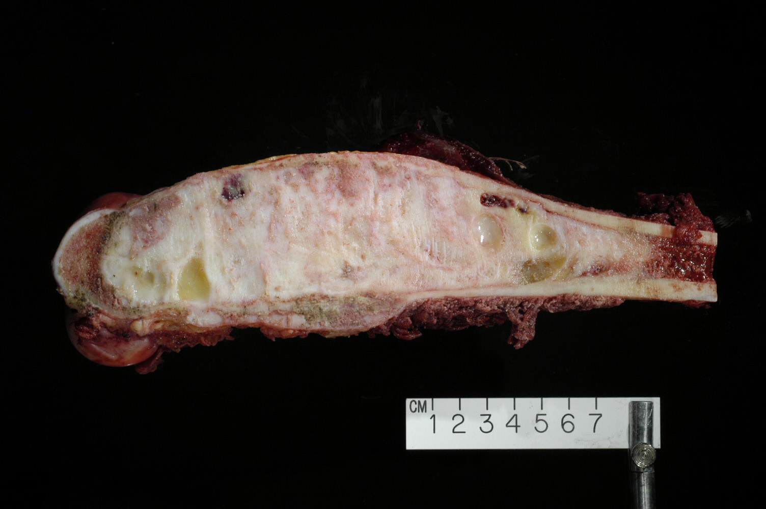
A 16 year old boy had a compound fracture of the right tibia after an apparent routine fall at a high-school soccer match. Radiology revealed a fracture through a lytic lesion of the proximal tibia with a "ground glass" appearance. During the repair of the fractured tibia, the surgeon curettaged the region of the lytic lesion and filled the defect with cement prior to internal stabilization of the fracture.
Gross Photo: [fibrous dysplasia of the tibia]
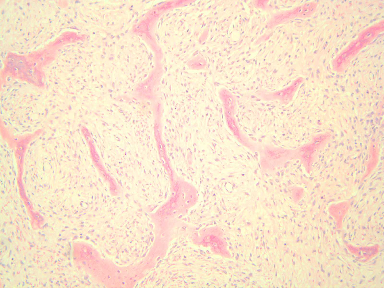
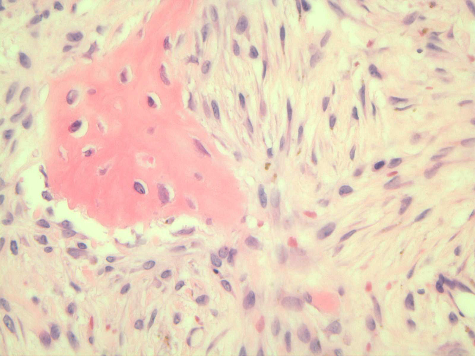
Photomicrograph: [fibrous dysplasia, intermediate and high power]
Case 2:
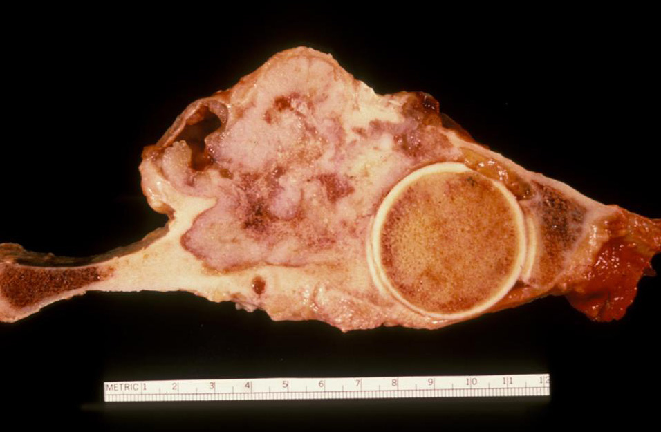
A 65 year old woman presented to her physician with severe pelvic pain. Radiology revealed a destructive lesion involving the bones of her pelvis extensively. A biopsy of the lesion was followed by a hemi-pelvectomy.
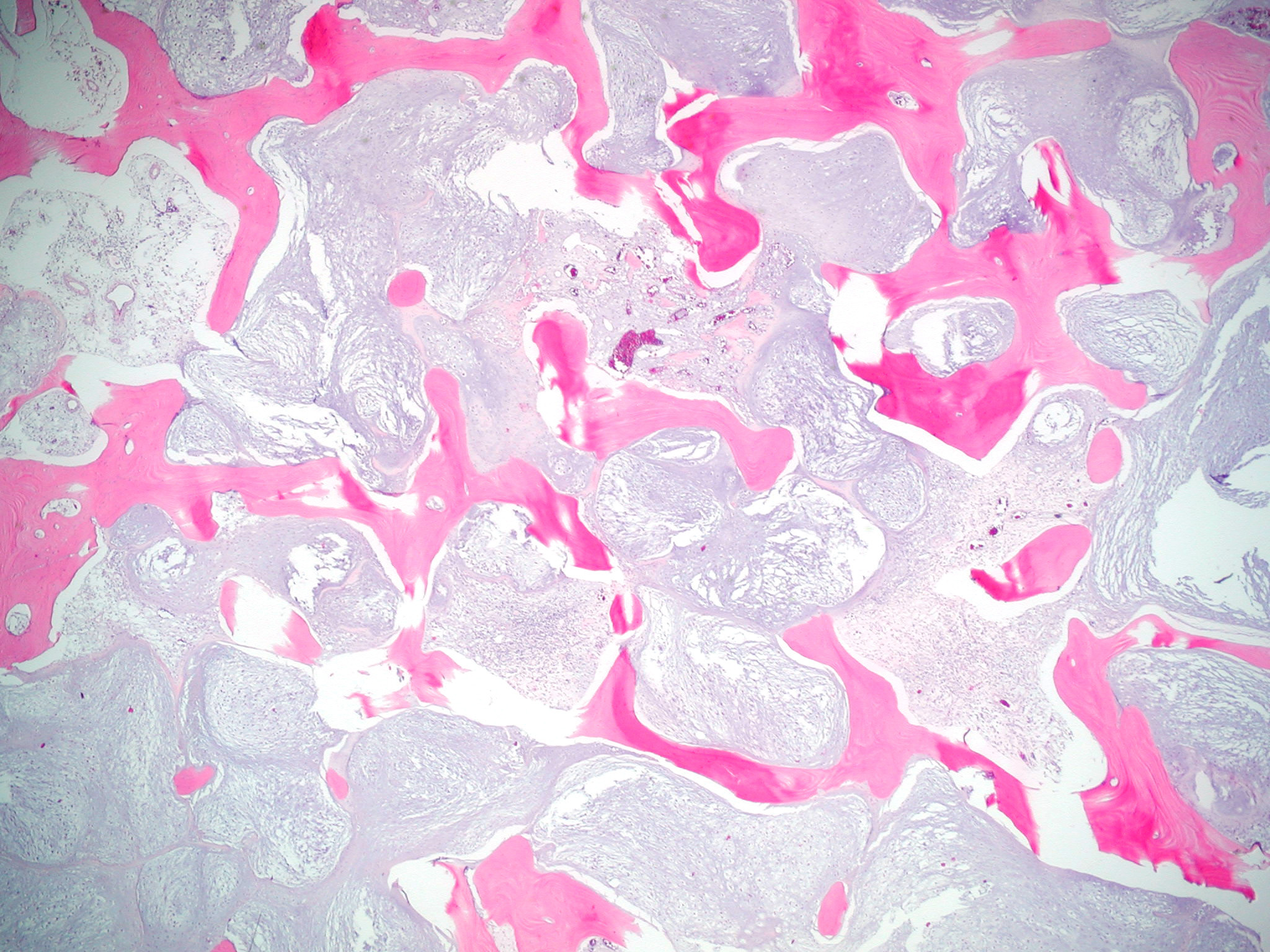
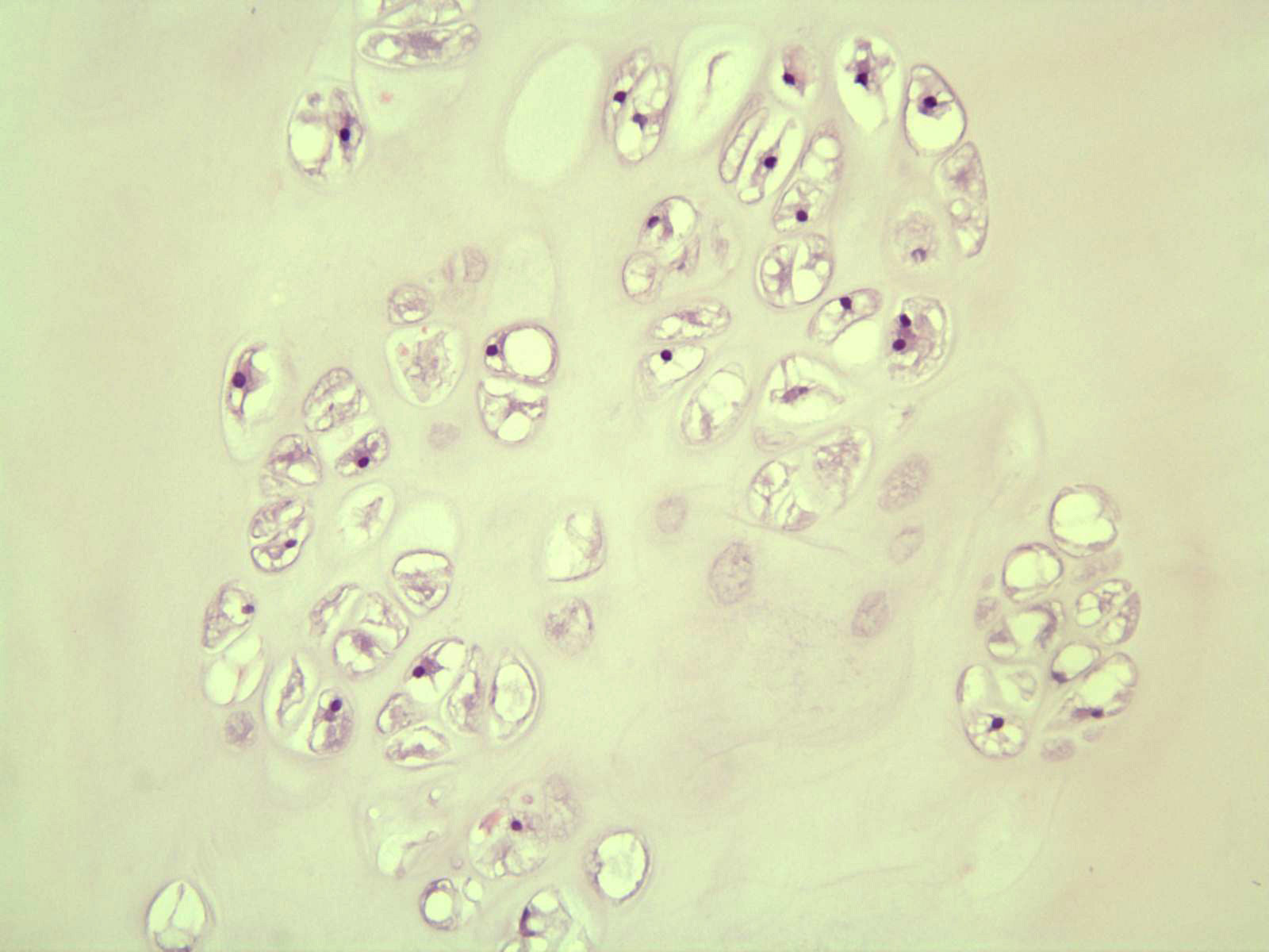
Gross Photo: [chondrosarcoma of the pelvis]
Photomicrograph: [chondrosarcoma showing permeative pattern]
Photomicrograph: [chondrosarcoma showing hypercellularity]
Case 3:
A 16 year old boy complained of right hip pain. Examination revealed an ill defined soft tissue mass in his upper right hip. Radiology showed a lytic lesion involving the proximal right femur with a large soft tissue component.
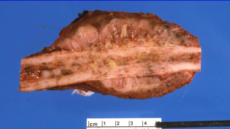
Gross Photo: [Ewing's sarcoma of the femur]
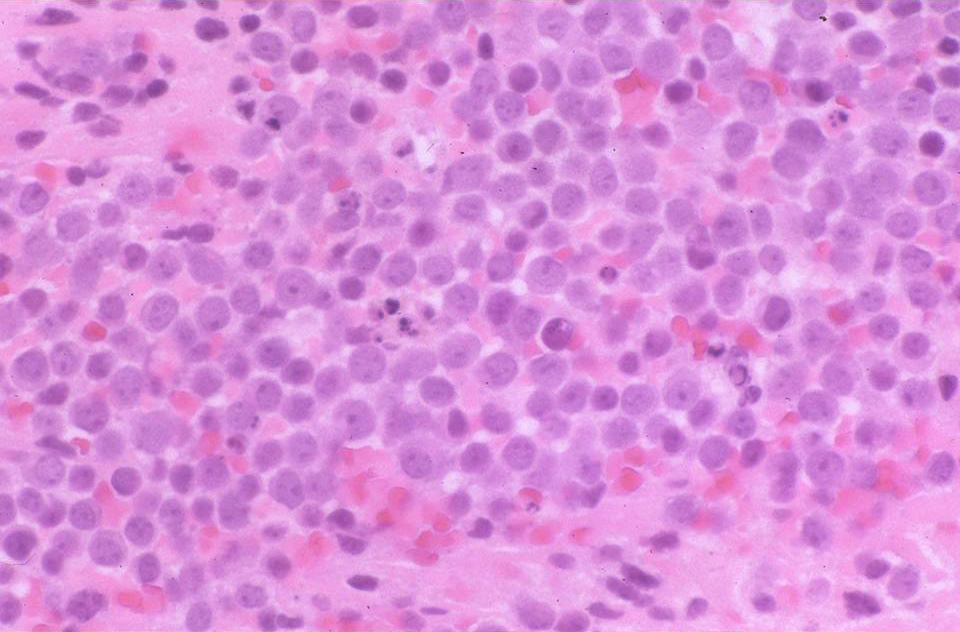
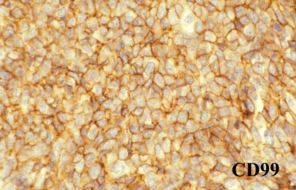
Photomicrograph: [Ewing's sarcoma]
Photomicrograph: [Ewing's sarcoma - immuno for CD99]
Case 4:
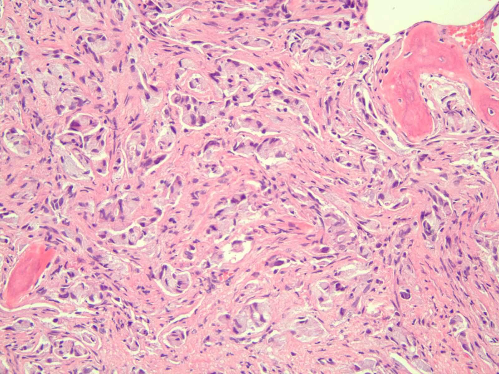
A 50 year old woman had unremitting pain of her right hip. A visit to her physician led to radiologic studies which showed a destructive lesion involving her femur.
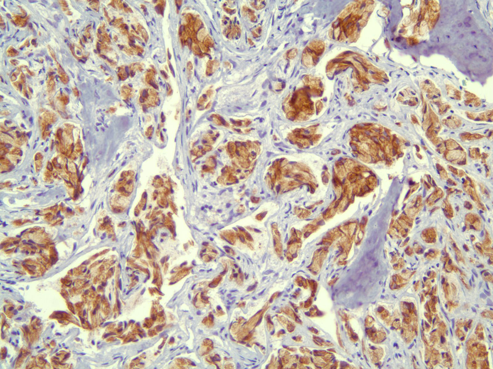
Photomicrograph: [metastatic adenocarcinoma of the lung]
Photomicrograph: [keratin immunohistochemistry]